Comparative Study
. 2001 Jun;75(11):4990-8.
doi: 10.1128/JVI.75.11.4990-4998.2001.

Identification of crucial hydrogen-bonding residues for the interaction of herpes simplex virus DNA polymerase subunits via peptide display, mutational, and calorimetric approaches

Affiliations
Comparative Study

Identification of crucial hydrogen-bonding residues for the interaction of herpes simplex virus DNA polymerase subunits via peptide display, mutational, and calorimetric approaches

K G Bridges et al. J Virol. 2001 Jun.

Abstract

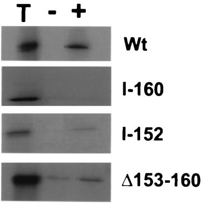
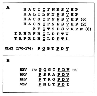
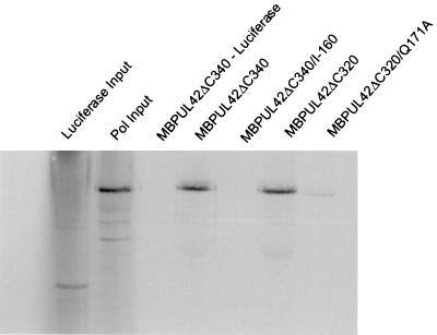
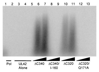
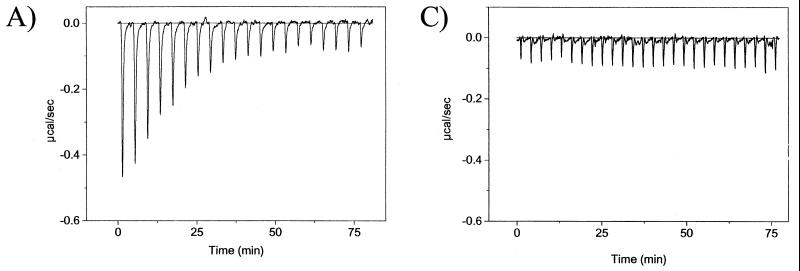
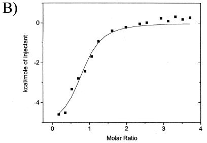
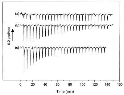
The catalytic subunit, Pol, of herpes simplex virus DNA polymerase interacts via its extreme C terminus with the processivity subunit, UL42. This interaction is critical for viral replication and thus a potential target for antiviral drug action. To investigate the Pol-binding region on UL42, we engineered UL42 mutations but also used random peptide display to identify artificial ligands of the Pol C terminus. The latter approach selected ligands with homology to residues 171 to 176 of UL42. Substitution of glutamine 171 with alanine greatly impaired binding to Pol and stimulation of long-chain DNA synthesis by Pol, identifying this residue as crucial for subunit interactions. To study these interactions quantitatively, we used isothermal titration calorimetry and wild-type and mutant forms of Pol-derived peptides and UL42. Each of three peptides corresponding to either the last 36, 27, or 18 residues of Pol bound specifically to UL42 in a 1:1 complex with a dissociation constant of 1 to 2 microM. Thus, the last 18 residues suffice for most of the binding energy, which was due mainly to a change in enthalpy. Substitutions at positions corresponding to Pol residue 1228 or 1229 or at UL42 residue 171 abolished or greatly reduced binding. These residues participate in hydrogen bonds observed in the crystal structure of the C terminus of Pol bound to UL42. Thus, interruption of these few bonds is sufficient to disrupt the interaction, suggesting that small molecules targeting the relevant side chains could interfere with Pol-UL42 binding.


Figures

FIG. 1
FIG. 1
Effects of mutations on binding UL42 to Pol. In vitro-expressed wt or mutant radiolabeled UL42 was incubated with either Pol (+) or β-Gal (−) and immunoprecipitated with antiserum directed against a Pol–β-Gal fusion, and the precipitates were analyzed by sodium dodecyl sulfate-polyacrylamide gel electrophoresis and autoradiography. Lane T, equivalent amount of input protein. At right are indicated the UL42 proteins assayed.
FIG. 2
FIG. 2
(A) Alignment of sequences of bacterial flagellar (top four) and phage (bottom three) display-derived peptides with residues 170 to 176 of UL42. The number of individual isolates is shown in parentheses. (Peptides whose sequences did not align are not shown.) (B) Alignment of residues 170 through 176 of UL42 with homologs from pseudorabies virus (PRV), equine herpesvirus 1 (EHV), and varicella-zoster virus (VZV).
FIG. 3
FIG. 3
MBP pulldown experiments. Radiolabeled wt or mutant MBP-UL42 proteins (as indicated at the top of the figure) were allowed to bind to amylose columns before loading in vitro-transcribed and translated, radiolabeled Pol or, where indicated, luciferase as a control. The columns were washed, and the proteins were eluted with 10 mM maltose. The first two lanes show 1/10th the amount of luciferase input and 1/15th the amount of Pol input, respectively. The following lanes show proteins eluted with maltose.
FIG. 4
FIG. 4
Ability of UL42 mutants to stimulate long-chain DNA synthesis by Pol. Experiments were performed using a poly(dA) template with an oligo(dT) primer and labeled TTP. The reaction products were visualized by autoradiography following electrophoresis on a 4% alkaline agarose gel. Lane 1,200 fmol of Pol alone; lanes 2 through 5,600 fmol of ΔC340, ΔC340/I-160, ΔC320, and ΔC320/Q171A alone, respectively. The remaining lanes contain 200 fmol of Pol plus 400 (lane 6) and 600 (lane 7) fmol of ΔC340, 400 (lane 8) and 600 (lane 9) fmol of ΔC340/I-160, 400 (lane 10) and 600 (lane 11) fmol of ΔC320, and 400 (lane 12) and 600 (lane 13) fmol of ΔC320/Q171A.
FIG. 5
FIG. 5
ITC of peptide A binding to UL42ΔC340. (A) Raw data for the titration of peptide A with ΔC340, in which the power output in microcalories per second is measured as a function of time in minutes. (B) The heats of dilution of both protein and ligand in A were subtracted, and the area under each injection curve was integrated to generate the points, which represent heat exchange in kilocalories per mole, which are plotted against the cumulative peptide-to-protein ratio for each injection. The solid line is the best-fit curve for the data. (C) Raw data for titration of peptide A with the I-160 mutant of UL42.
FIG. 5
FIG. 5
ITC of peptide A binding to UL42ΔC340. (A) Raw data for the titration of peptide A with ΔC340, in which the power output in microcalories per second is measured as a function of time in minutes. (B) The heats of dilution of both protein and ligand in A were subtracted, and the area under each injection curve was integrated to generate the points, which represent heat exchange in kilocalories per mole, which are plotted against the cumulative peptide-to-protein ratio for each injection. The solid line is the best-fit curve for the data. (C) Raw data for titration of peptide A with the I-160 mutant of UL42.
FIG. 6
FIG. 6
CD spectra of peptides A and G. Wavelength scans of peptides in 10 mM KF were recorded at 1-nm intervals with a 1-s averaging time, and 10 to 15 scans were averaged. ⧫, peptide A; ■, peptide G.
FIG. 7
FIG. 7
ITC of H1228 and R1229 variants of peptide E. Raw data are shown as in Fig. 1A for the interaction of (a) H1228A, (b) R1229A, and (c) wt peptide E with UL42ΔC340. Experiments were performed using 7.2 μM UL42ΔC340 and 335 μM peptide. The arrow indicates the scale for heat exchange in microcalories per second.

References

    1. Ausubel F M, Brent R, Kingston R E, Moore D D, Seidman J G, Smith J A, Struhl K, editors. Current protocols in molecular biology. New York, N.Y: Greene Publishing Associates and Wiley Interscience; 1987.
    1. Berthomme H, Monahan S J, Parris D S, Jacquemont B, Epstein A L. Cloning, sequencing, and functional characterization of the two subunits of the pseudorabies virus DNA polymerase holoenzyme: evidence for specificity of interaction. J Virol. 1995;69:2811–2818. - PMC - PubMed
    1. Bridges K G, Hua Q, Brigham-Burke M R, Martin J D, Hensley P, Dahl C E, Weiss M A, Coen D M. Secondary structure and structure-activity relationships of peptides corresponding to the subunit interface of herpes simplex virus DNA polymerase. J Biol Chem. 2000;275:472–478. - PubMed
    1. Chow C S, Coen D M. Mutations that specifically impair the DNA binding activity of the herpes simplex virus protein UL42. J Virol. 1995;69:6965–6971. - PMC - PubMed
    1. Digard P, Bebrin W R, Weisshart K, Coen D M. The extreme C terminus of herpes simplex virus DNA polymerase is crucial for functional interaction with processivity factor UL42 and for viral replication. J Virol. 1993;67:398–406. - PMC - PubMed

Publication types

LinkOut - more resources