Comparative Study
. 2001 Jun;75(11):5018-26.
doi: 10.1128/JVI.75.11.5018-5026.2001.

Infectious entry by amphotropic as well as ecotropic murine leukemia viruses occurs through an endocytic pathway

Affiliations
Comparative Study

Infectious entry by amphotropic as well as ecotropic murine leukemia viruses occurs through an endocytic pathway

L J Katen et al. J Virol. 2001 Jun.

Abstract

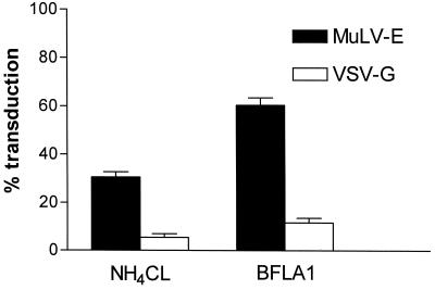
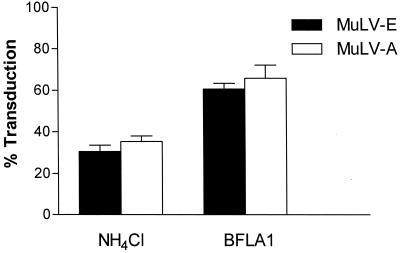
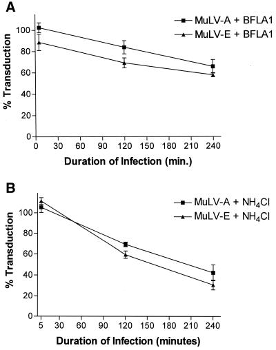
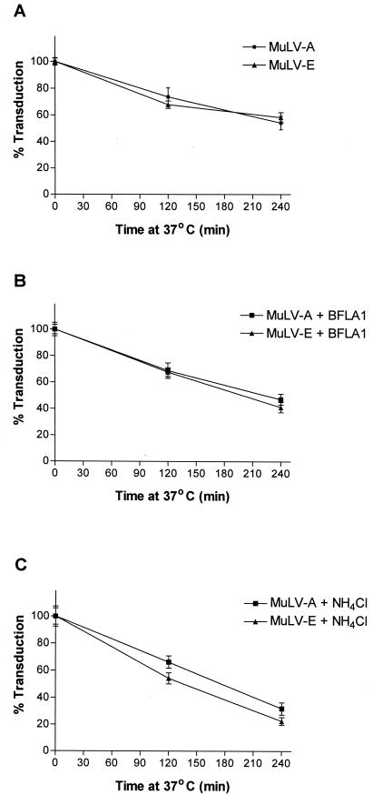
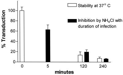
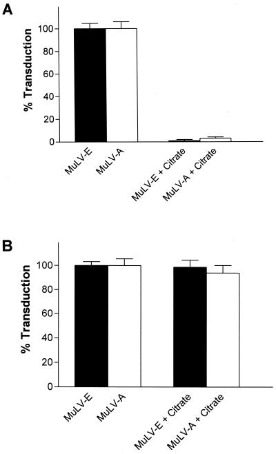
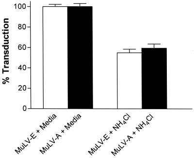
Infectious entry of enveloped viruses is thought to proceed by one of two mechanisms. pH-dependent viruses enter the cells by receptor-mediated endocytosis and are inhibited by transient treatment with agents that prevent acidification of vesicles in the endocytic pathway, while pH-independent viruses are not inhibited by such agents and are thought to enter the cell by direct fusion with the plasma membrane. Nearly all retroviruses, including amphotropic murine leukemia virus (MuLV) and human immunodeficiency virus type 1, are classified as pH independent. However, ecotropic MuLV is considered to be a pH-dependent virus. We have examined the infectious entry of ecotropic and amphotropic MuLVs and found that they were equally inhibited by NH4Cl and bafilomycin A. These agents inhibited both viruses only partially over the course of the experiments. Agents that block the acidification of endocytic vesicles also arrest vesicular trafficking. Thus, partial inhibition of the MuLVs could be the result of virus inactivation during arrest in this pathway. In support of this contention, we found that that the loss of infectivity of the MuLVs during treatment of target cells with the drugs closely corresponded to the loss of activity due to spontaneous inactivation at 37 degrees C in the same period of time. Furthermore, the drugs had no effect on the efficiency of infection under conditions in which the duration of infection was held to a very short period to minimize the effects of spontaneous inactivation. These results indicate that the infectious processes of both ecotropic and amphotropic MuLVs were arrested rather than aborted by transient treatment of the cells with the drugs. We also found that infectious viruses were efficiently internalized during treatment. This indicated that the arrest occurred in an intracellular compartment and that the infectious process of both the amphotropic and ecotropic MuLVs very likely involved endocytosis. An important aspect of this study pertains to the interpretation of experiments in which agents that block endocytic acidification inhibit infectivity. As we have found with the MuLVs, inhibition of infectivity may be secondary to the block of endocytic acidification. While this strongly suggests the involvement of an endocytic pathway, it does not necessarily indicate a requirement for an acidic compartment during the infectious process. Likewise, a lack of inhibition during transient treatment with the drugs would not preclude an endocytic pathway for viruses that are stable during the course of the treatment.


Figures

FIG. 1
FIG. 1
Partial inhibition of MuLV-E transduction by NH4Cl or BFLA1. Cells were incubated for 30 min in the presence of NH4Cl or BFLA1 and subsequently infected with a mixture of LAPSN(MuLV-E) and G1nβgSvNa(VSV-G) in medium containing either drug for a period of 2 h. The drug-virus mixture was then replaced by medium containing the drug and incubated for an additional 2 h. The medium containing the drug was then replaced by fresh medium; the culture was allowed to grow to confluence and assayed for transduction by the retroviral vectors as described in Materials and Methods. Percentages of transduction were calculated from the mean values of parallel assays performed in the absence of the drugs. Data for cells treated with NH4Cl represent the means and SEM of 12 determinations in two separate experiments; data for cells treated by BFLA1 represent the means and SEM of 10 determinations in two separate experiments.
FIG. 2
FIG. 2
Inhibition of MuLV-E and MuLV-A by NH4Cl or BFLA1. Cells were treated with the drugs and infected with a mixture of LAPSN(MuLV-E) and G1nβgSvNa(MuLV-A) in the presence of the drugs as described in the legend to Fig. 1. Data for cells treated with NH4Cl represent the means and SEM of 21 determinations in four separate experiments. Percentages of transduction were calculated from the mean values of parallel assays performed in the absence of the drugs. Data for cells treated with BFLA1 represent the means and SEM of 22 determinations in four separate experiments.
FIG. 3
FIG. 3
Duration of infection and inhibition of MuLV-E and MuLV-A by BFLA1 (A) or NH4Cl (B). Cells were preincubated with medium containing the drugs and infected with a mixture of LAPSN(MuLV-E) and GlnβgSvNa(MuLV-A) in the presence of the drugs after different times of preincubation. All cultures were exposed to the drugs for a period of 4.5 h. Preincubation times were 30 min, 2.5 h, and 4 h 25 min such that the duration of infection were 4 h, 2 h, and 5 min, respectively, before removal of the drug. Percentages of transduction were calculated from the mean values of parallel assays performed in the absence of the drugs. Each data point for cells treated with NH4Cl represents the mean and SEM of 13 to 22 determination in three to four separate experiments; each data point for cells treated with BFLA1 represents the mean and SEM of 11 to 22 determination in three to four separate experiments.
FIG. 4
FIG. 4
Spontaneous inactivation of MuLV-E and MuLV-A in the presence of NH4Cl or BFLA1. A mixture of LAPSN(MuLV-E) and G1nβgSvNa(MuLV-A) was incubated in a water bath at 37°C in medium or in medium containing 0.05 μM BFLA1 or 50 mM NH4Cl. Aliquots of the virus mixture were removed at 0, 2, and 4 h and assayed for transduction activity as described in Materials and Methods. Mean transduction titers obtained after 0 h of incubation were considered to be 100%, and percent transduction after 2 and 4 h was calculated relative to the mean titers obtained at 0 h. Data for virus incubated in medium (A), medium with BFLA1 (B), and medium with NH4Cl (C) represent the means and SEM of 15 determinations at each time in four separate experiments.
FIG. 5
FIG. 5
Correlation of spontaneous inactivation with the inhibition of VSV-G by NH4Cl. To determine the effect of the duration of infection on inhibition by NH4Cl, cells were preincubated in medium containing NH4Cl and infected with G1nβgSvNa(VSV-G) in the presence of NH4Cl after different times of preincubation as described for Fig. 3. The data represent the mean and SEM of 9 to 15 determinations at each time in two to four separate experiments. For thermal stability, G1nβgSvNa(VSV-G) was incubated in a water bath at 37°C in the presence of NH4Cl. Aliquots of the virus mixture were removed at 0, 2, and 4 h and assayed for transduction activity as described in Materials and Methods. Mean transduction titers obtained after 0-h incubation were considered to be 100%, and percent transduction after 2 and 4 h was calculated relative to the mean titers obtained at 0 h. Each value represents the mean and SEM of six determinations at each time in two separate experiments.
FIG. 6
FIG. 6
Inactivation of cell surface-bound MuLV-E and MuLV-A by treatment with citrate buffer (pH 3.0). (A) Cells were incubated for 2 h at 4°C with a mixture of LAPSN(MuLV-E) and G1nβgSvNa(MuLV-A); the cells washed and then mock treated or treated with citrate buffer (pH 3) as described in Materials and Methods. Each value represents the mean and SEM of 12 determinations in two separate experiments. (B) Cells were mock treated or treated with citrate buffer (pH 3) as described in the text. Immediately after treatment the cells were infected for 2 h at 37°C with a mixture of LAPSN(MuLV-E) and G1nβgSvNa(MuLV-A). Each value represents the mean and SEM of eight determinations in two separate experiments.
FIG. 7
FIG. 7
Effect of NH4Cl on entry of MuLV-E and MuLV-A. Cells were incubated for 30 min at 37°C in medium or medium containing NH4Cl and then infected with a mixture of LAPSN(MuLV-E) and G1nβgSvNa(MuLV-A) for 2 h in the presence or absence of the base. The cells were then treated with citrate buffer (pH 3) as described in the text. Each value represents the mean and SEM of 10 determinations in three separate experiments.

Similar articles

Cited by

References

    1. Aiken C. Pseudotyping human immunodeficiency virus type 1 (HIV-1) by the glycoprotein of vesicular stomatitis virus targets HIV-1 entry to an endocytic pathway and suppresses both the requirement for Nef and the sensitivity to cyclosporin A. J Virol. 1997;71:5871–5877. - PMC - PubMed
    1. Andersen K B, Nexo B A. Entry of murine retrovirus into mouse fibroblasts. Virology. 1983;125:85–98. - PubMed
    1. Andreadis S, Palsson B O. Coupled effects of polybrene and calf serum on the efficiency of retroviral transduction and the stability of retroviral vectors. Hum Gene Ther. 1997;8:285–291. - PubMed
    1. Andreadis S T, Brott D, Fuller A O, Palsson B O. Moloney murine leukemia virus-derived retroviral vectors decay intracellularly with a half-life in the range of 5.5 to 7.5 hours. J Virol. 1997;71:7541–7548. - PMC - PubMed
    1. Bayner N, Schober D, Prchla E, Murphy R F, Blaas D, Fuchs R. Effect of bafilomycin A1 and nocodazole on endocytic transport in HeLa cells: implication for viral uncoating and infection. J Virol. 1998;72:9645–9655. - PMC - PubMed

Publication types