Incorporation of lysyl-tRNA synthetase into human immunodeficiency virus type 1
- PMID: 11333884
- PMCID: PMC114908
- DOI: 10.1128/JVI.75.11.5043-5048.2001
Incorporation of lysyl-tRNA synthetase into human immunodeficiency virus type 1
Abstract
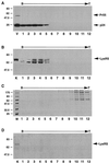
During human immunodeficiency virus type 1 (HIV-1) assembly, tRNA(Lys) isoacceptors are selectively incorporated into virions and tRNA(Lys)3 is used as the primer for reverse transcription. We show herein that the tRNA(Lys)-binding protein, lysyl-tRNA synthetase (LysRS), is also selectively packaged into HIV-1. The viral precursor protein Pr55gag alone will package LysRS into Pr55gag particles, independently of tRNA(Lys). With the additional presence of the viral precursor protein Pr160gag-pol, tRNA(Lys) and LysRS are both packaged into the particle. While the predominant cytoplasmic LysRS has an apparent M(r) of 70,000, viral LysRS associated with tRNA(Lys) packaging is shorter, with an apparent M(r) of 63,000. The truncation occurs independently of viral protease and might be required to facilitate interactions involved in the selective packaging and genomic placement of primer tRNA.
Figures


Similar articles
-
Ability of wild-type and mutant lysyl-tRNA synthetase to facilitate tRNA(Lys) incorporation into human immunodeficiency virus type 1.J Virol. 2004 Feb;78(3):1595-601. doi: 10.1128/jvi.78.3.1595-1601.2004. J Virol. 2004. PMID: 14722314 Free PMC article.
-
Formation of the tRNALys packaging complex in HIV-1.FEBS Lett. 2010 Jan 21;584(2):359-65. doi: 10.1016/j.febslet.2009.11.038. FEBS Lett. 2010. PMID: 19914238 Free PMC article. Review.
-
Role of Pr160gag-pol in mediating the selective incorporation of tRNA(Lys) into human immunodeficiency virus type 1 particles.J Virol. 1994 Apr;68(4):2065-72. doi: 10.1128/JVI.68.4.2065-2072.1994. J Virol. 1994. PMID: 7511167 Free PMC article.
-
Specific inhibition of the synthesis of human lysyl-tRNA synthetase results in decreases in tRNA(Lys) incorporation, tRNA(3)(Lys) annealing to viral RNA, and viral infectivity in human immunodeficiency virus type 1.J Virol. 2003 Sep;77(18):9817-22. doi: 10.1128/jvi.77.18.9817-9822.2003. J Virol. 2003. PMID: 12941890 Free PMC article.
-
The tRNALys packaging complex in HIV-1.Int J Biochem Cell Biol. 2004 Sep;36(9):1776-86. doi: 10.1016/j.biocel.2004.02.022. Int J Biochem Cell Biol. 2004. PMID: 15183344 Review.
Cited by
-
Reverse Transcriptase and Cellular Factors: Regulators of HIV-1 Reverse Transcription.Viruses. 2009 Dec;1(3):873-94. doi: 10.3390/v1030873. Epub 2009 Nov 10. Viruses. 2009. PMID: 21994574 Free PMC article.
-
Binding of the eukaryotic translation elongation factor 1A with the 5'UTR of HIV-1 genomic RNA is important for reverse transcription.Virol J. 2015 Aug 6;12:118. doi: 10.1186/s12985-015-0337-x. Virol J. 2015. PMID: 26242867 Free PMC article.
-
Profiling non-lysyl tRNAs in HIV-1.RNA. 2010 Feb;16(2):267-73. doi: 10.1261/rna.1928110. Epub 2009 Dec 9. RNA. 2010. PMID: 20007329 Free PMC article.
-
HIV Genome-Wide Protein Associations: a Review of 30 Years of Research.Microbiol Mol Biol Rev. 2016 Jun 29;80(3):679-731. doi: 10.1128/MMBR.00065-15. Print 2016 Sep. Microbiol Mol Biol Rev. 2016. PMID: 27357278 Free PMC article.
-
Effect of Lysyl-tRNA Synthetase on the Maturation of HIV-1 Reverse Transcriptase.ACS Omega. 2020 Jun 30;5(27):16619-16627. doi: 10.1021/acsomega.0c01449. eCollection 2020 Jul 14. ACS Omega. 2020. PMID: 32685828 Free PMC article.
References
-
- Berkowitz R, Fisher J, Goff S P. RNA packaging. In: Krausslich H G, editor. Morphogenesis and maturation of retroviruses. Vol. 214. New York, N.Y: Springer-Verlag; 1996. pp. 177–218. - PubMed
-
- Bradford M M. A rapid and sensitive method for the quantitation of microgram quantities of protein utilizing the principle of protein-dye binding. Anal Biochem. 1976;72:248–254. - PubMed
MeSH terms
Substances
LinkOut - more resources
Full Text Sources
Other Literature Sources

