

Herpes simplex virus type 1 corneal infection results in periocular disease by zosteriform spread
- PMID: 11333887
- PMCID: PMC114911
- DOI: 10.1128/JVI.75.11.5069-5075.2001
Herpes simplex virus type 1 corneal infection results in periocular disease by zosteriform spread
Abstract
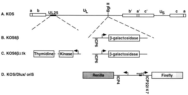
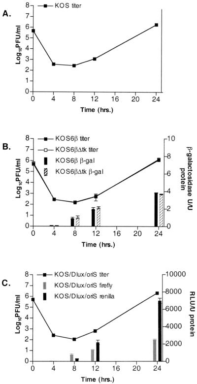
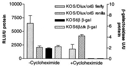
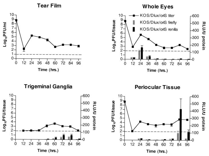
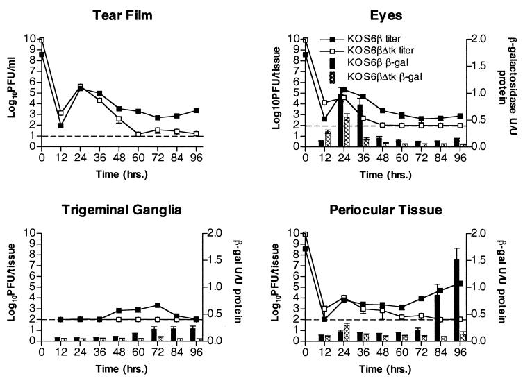
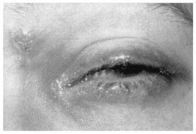
In humans and animal models of herpes simplex virus infection, zosteriform skin lesions have been described which result from anterograde spread of the virus following invasion of the nervous system. Such routes of viral spread have not been fully examined following corneal infection, and the possible pathologic consequences of such spread are unknown. To investigate this, recombinant viruses expressing reporter genes were generated to quantify and correlate gene expression with replication in eyes, trigeminal ganglia, and periocular tissue. Reporter activity peaked in eyes 24 h postinfection and rapidly fell to background levels by 48 h despite the continued presence of viral titers. Reporter activity rose in the trigeminal ganglia at 60 h and peaked at 72 h, concomitant with the appearance and persistence of infectious virus. Virus was present in the periocular skin from 24 h despite the lack of significant reporter activity until 84 h postinfection. This detection of reporter activity was followed by the onset of periocular disease on day 4. Corneal infection with a thymidine kinase-deleted reporter virus displayed a similar profile of reporter activity and viral titer in the eyes, but little or no detectable activity was observed in trigeminal ganglia or periocular tissue. In addition, no periocular disease symptoms were observed. These findings demonstrate that viral infection of periocular tissue and subsequent disease development occurs by zosteriform spread from the cornea to the periocular tissue via the trigeminal ganglion rather than by direct spread from cornea to the periocular skin. Furthermore, clinical evidence is discussed suggesting that a similar mode of spreading and disease occurs in humans following primary ocular infection.
Figures


Similar articles
-
A replication competent HSV-1(McKrae) with a mutation in the amino-terminus of glycoprotein K (gK) is unable to infect mouse trigeminal ganglia after cornea infection.Curr Eye Res. 2014 Jun;39(6):596-603. doi: 10.3109/02713683.2013.855238. Epub 2014 Jan 8. Curr Eye Res. 2014. PMID: 24401006
-
Importance of the herpes simplex virus UL24 gene for productive ganglionic infection in mice.Virology. 1998 Mar 1;242(1):161-9. doi: 10.1006/viro.1997.9012. Virology. 1998. PMID: 9501052
-
Tracking the spread of a lacZ-tagged herpes simplex virus type 1 between the eye and the nervous system of the mouse: comparison of primary and recurrent infection.J Virol. 2001 Jun;75(11):5252-62. doi: 10.1128/JVI.75.11.5252-5262.2001. J Virol. 2001. PMID: 11333907 Free PMC article.
-
Type I interferon and lymphangiogenesis in the HSV-1 infected cornea - are they beneficial to the host?Prog Retin Eye Res. 2013 Sep;36:281-91. doi: 10.1016/j.preteyeres.2013.06.003. Epub 2013 Jul 19. Prog Retin Eye Res. 2013. PMID: 23876483 Free PMC article. Review.
-
Periocular dermatoses.Int J Womens Dermatol. 2017 Sep 18;3(4):206-218. doi: 10.1016/j.ijwd.2017.08.001. eCollection 2017 Dec. Int J Womens Dermatol. 2017. PMID: 29234715 Free PMC article. Review.
Cited by
-
Artemisinin-derived dimer diphenyl phosphate is an irreversible inhibitor of human cytomegalovirus replication.Antimicrob Agents Chemother. 2012 Jul;56(7):3508-15. doi: 10.1128/AAC.00519-12. Epub 2012 Apr 30. Antimicrob Agents Chemother. 2012. PMID: 22547612 Free PMC article.
-
Complement Suppresses the Initial Type 1 Interferon Response to Ocular Herpes Simplex Virus Type 1 Infection in Mice.Pathogens. 2024 Jan 13;13(1):74. doi: 10.3390/pathogens13010074. Pathogens. 2024. PMID: 38251381 Free PMC article.
-
Regulation of the catalytic activity of herpes simplex virus 1 protein kinase Us3 by autophosphorylation and its role in pathogenesis.J Virol. 2009 Jun;83(11):5773-83. doi: 10.1128/JVI.00103-09. Epub 2009 Mar 18. J Virol. 2009. PMID: 19297494 Free PMC article.
-
Herpesvirus entry mediator is a serotype specific determinant of pathogenesis in ocular herpes.Proc Natl Acad Sci U S A. 2012 Dec 11;109(50):20649-54. doi: 10.1073/pnas.1216967109. Epub 2012 Nov 26. Proc Natl Acad Sci U S A. 2012. PMID: 23184983 Free PMC article.
-
Lytic Promoters Express Protein during Herpes Simplex Virus Latency.PLoS Pathog. 2016 Jun 27;12(6):e1005729. doi: 10.1371/journal.ppat.1005729. eCollection 2016 Jun. PLoS Pathog. 2016. PMID: 27348812 Free PMC article.
References
-
- Adams R, Cunningham C, Davison M D, MacLean C A, Davison A J. Characterization of the protein encoded by gene UL49A of herpes simplex virus type 1. J Gen Virol. 1998;79:813–823. - PubMed
-
- Awan A R, Harmenberg J, Flink O, Field H J. Combinations of antiviral and anti-inflammatory preparations for the topical treatment of herpes simplex virus assessed using a murine zosteriform infection model. Antivir Chem Chemother. 1998;9:19–24. - PubMed
-
- Bloom D C, Jarman R G. Generation and use of recombinant reporter viruses for study of herpes simplex virus infections in vivo. Methods. 1998;16:117–125. - PubMed
-
- Blyth W A, Harbour D A, Hill T J. Pathogenesis of zosteriform spread of herpes simplex virus in the mouse. J Gen Virol. 1984;65:1477–1486. - PubMed
-
- Brandt C R, Kintner R L, Pumfery A M, Visalli R J, Grau D R. The herpes simplex virus ribonucleotide reductase is required for ocular virulence. J Gen Virol. 1991;72:2043–2049. - PubMed
Publication types
MeSH terms
Substances
Grants and funding
LinkOut - more resources
Full Text Sources
Other Literature Sources
Medical
