Comparative Study
. 2001 Jun;75(11):5159-73.
doi: 10.1128/JVI.75.11.5159-5173.2001.

Marek's disease virus (MDV) encodes an interleukin-8 homolog (vIL-8): characterization of the vIL-8 protein and a vIL-8 deletion mutant MDV

Affiliations
Free PMC article
Comparative Study

Marek's disease virus (MDV) encodes an interleukin-8 homolog (vIL-8): characterization of the vIL-8 protein and a vIL-8 deletion mutant MDV

M S Parcells et al. J Virol. 2001 Jun.
Free PMC article

Abstract

Chemokines induce chemotaxis, cell migration, and inflammatory responses. We report the identification of an interleukin-8 (IL-8) homolog, termed vIL-8, encoded within the genome of Marek's disease virus (MDV). The 134-amino-acid vIL-8 shares closest homology to mammalian and avian IL-8, molecules representing the prototype CXC chemokine. The gene for vIL-8 consists of three exons which map to the BamHI-L fragment within the repeats flanking the unique long region of the MDV genome. A 0.7-kb transcript encoding vIL-8 was detected in an n-butyrate-treated, MDV-transformed T-lymphoblastoid cell line, MSB-1. This induction is essentially abolished by cycloheximide and herpesvirus DNA polymerase inhibitor phosphonoacetate, indicating that vIL-8 is expressed with true late (gamma2) kinetics. Baculovirus-expressed vIL-8 was found to be secreted into the medium and shown to be functional as a chemoattractant for chicken peripheral blood mononuclear cells but not for heterophils. To characterize the function of vIL-8 with respect to MDV infection in vivo, a recombinant MDV was constructed with a deletion of all three exons and a soluble-modified green fluorescent protein (smGFP) expression cassette inserted at the site of deletion. In two in vivo experiments, the vIL-8 deletion mutant (RB1BvIL-8DeltasmGFP) showed a decreased level of lytic infection in comparison to its parent virus, an equal-passage-level parent virus, and to another recombinant MDV containing the insertion of a GFP expression cassette at the nonessential US2 gene. RB1BvIL-8DeltasmGFP retained oncogenicity, albeit at a greatly reduced level. Nonetheless, we have been able to establish a lymphoblastoid cell line from an RB1BvIL-8DeltasmGFP-induced ovarian lymphoma (MDCC-UA20) and verify the presence of a latent MDV genome lacking vIL-8. Taken together, these data describe the identification and characterization of a chemokine homolog encoded within the MDV genome that is dispensable for transformation but may affect the level of MDV in vivo lytic infection.


Figures
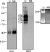
FIG. 1
FIG. 1
Expression of vIL-8 is dramatically induced in MSB-1 cells treated with n-butyrate. For analysis, 4 × 10⁶ MSB-1 cells were not treated (U) or treated with 3 mM n-butyrate (NB) for 48 h at 37°C to induce virus replication. Total RNA was purified from each sample and subjected to Northern blot hybridization analysis. Duplicate blots were prepared for different hybridizations. A distinct 0.7-kb transcript was identified when a DNA fragment containing vIL-8 sequences was used as a probe (vIL8). Without NB treatment, at least five transcripts could be identified using a probe encompassing the latent MDV EcoRI Q protein Meq (MEQ). As a comparison, chicken cellular rRNAs (28S and 18S) were stained with ethidium bromide to demonstrate the cellular transcription level in MSB-1 before and after NB treatment.
FIG. 2
FIG. 2
Characterization of transcript encoding vIL-8 by RACE analysis. (A) Schematic drawing of the MDV genome and the location of vIL-8. Transcripts encompassing meq, meqΔC-BamL, and vIL-8 are depicted at the bottom of the diagram with exons in boxes and introns in solid lines. Abbreviations: IRL, long internal repeat; IRS, short internal repeat; TRL, long terminal repeat; TRS, short terminal repeat; UL, unique long sequence; US, unique short sequence. (B) Locations of the oligonucleotide primers used in RACE reactions. vIL-8F4 was used as a gene-specific primer in 3′ RACE. vIL-8B1 was used as a gene-specific primer in 5′ RACE. vIL-8FL-S and vIL-8FL-AS are primers derived from RACE results used to amplify the full length of the vIL-8 open reading frame. The empty arrows depict exons II and III present in the meqΔC-BamL and vIL-8 sequences. (C) Ethidium bromide-stained agarose gel (1%) demonstrates 0.3- and 0.45-kb PCR amplicons in 5′- and 3′ RACE reactions, respectively. Sizes are given on left in kilobases.
FIG. 3
FIG. 3
Fine structure of vIL-8 cDNA. (A) The nucleotide sequence of a full-length cDNA compared to its genomic counterpart. cDNA sequences are in capitals and blocked by lines while consensus splice donor/splice acceptor in introns are in bold. Putative TATA box (TATATAA) and polyadenylation recognition (ATTAAA) sequences are underlined. Deduced amino acid sequences are shown beneath the nucleotide sequences. The 17 hydrophobic residues of the first exon are in boldface. Four cysteines conserved in all CXC chemokines are circled. (B) Alignment of amino acid sequences of vIL-8 and various mammalian and avian IL-8 homologs. The four conserved cysteines and the signature ELR motif are indicated on top of the alignment. Similar (shaded in light gray) and identical (shaded in dark gray) amino acids are indicated. The abbreviation used and GenBank accession number for each sequence are rhIL8 (rhesus macaque IL-8), P51495; huIL8 (hIL-8), P10145; ovIL8 (ovine IL-8), P36925; doIL8 (dog IL-8), JN0841; rabIL8 (rabbit IL-8), P19874; 9E3/CEF4 (CEF protein 9E3), M16199; moGro (mouse Gro), P12850; moMIP2 (murine macrophage inflammatory protein 2), P10889; moBLC (Mus musculus B-lymphocyte chemoattractant), AF044196; and ratPF4 (rat PF4), P06765.
FIG. 4
FIG. 4
Expression kinetics of vIL-8 in MSB-1 cells. (A) MSB-1 cells were treated with n-butyrate (3 mM) for 0 to 48 h, as indicated. For the 36-h time point, duplicate cell cultures were prepared for the treatment of cells with PAA (100 μM). Total RNAs were extracted at the end of chemical treatment, and concentrations were determined. Twenty micrograms of RNA from each sample was loaded to each lane, and duplicate filters were prepared for vIL-8 hybridization (upper gel) and GAPDH hybridization (lower gel) (33). (B) RNA was purified from RB1B-infected CEF that were not treated (left lanes), treated with 100 μg of CHX/ml (center lanes), or treated with 100 μg of PAA/ml (right lanes) and was used as the template for RT-PCR. For each RNA treatment group, three concentrations of RNA were used (500, 50 and 5 ng, left to right, respectively). RT-PCR was performed as described in the text, and the entire reaction products (total, 25 μl) were loaded on a 1% agarose gel. The products for RT-PCR were ICP27, 670 bp; RRL, 549 bp; gC, 584 bp; vIL-8, 699 bp (unspliced) and 423 bp (spliced); and Meq, 421 bp (Table 1). Arrows indicate the RT-PCR products of the appropriate size. Far right lanes contain molecular size markers (Hyper-Ladder; Bioline). (C) Ethidium bromide-stained agarose gel of 1 μg of each RNA used for RT-PCR analysis in panel B, demonstrating that the RNAs used were not degraded. Mrk, Hyper-Ladder DNA size markers in base pairs. Un, untreated.
FIG. 5
FIG. 5
Purification of six-His-tagged vIL-8 produced by baculovirus expression system. (A) Purification of recombinant vIL-8–His by Ni-affinity column chromatography. Cell-free supernatants collected from High Five insect cells infected with vIL-8–His recombinant baculovirus were prepared and loaded onto a Ni column as described in Materials and Methods. Bound vIL-8–His was eluted by a 20 to 500 mM imidazole gradient. Twenty microliters of each elution fraction was loaded onto an SDS–10 to 20% PAGE gel, separated electrophoretically, and stained with Coomassie blue. Kd, kilodaltons. (B) Silver staining demonstrates the purity of vIL-8–His after gel filtration. Elution fractions 3 and 4 of the Ni column were pooled, concentrated eightfold by Centricon-3 spin concentration, and further purified by gel filtration chromatography (Superose 12). Fractions were collected every 2 min at a flow rate of 0.4 min/ml. Purity of the eluent with the highest chemotaxis activity (42- to 44-min fractions) was analyzed by SDS–10 to 20% PAGE followed by silver staining. A major protein band with a size of approximately 20 kDa is indicated with an asterisk.
FIG. 6
FIG. 6
Chemotaxis assays demonstrate that MDV vIL-8 is a functional chicken PBMC chemoattractant. The chemotactic activity of vIL-8 toward chicken PBMC (A) or heterophils (B) was assayed using Costar Transwell plates. For panel A, chemotaxis was performed using 5-μm-pore-size Transwell plates with the indicated concentrations of vIL-8 at 37°C for 40 min. For panel B, a 3-μm-pore-size Transwell plate was used for heterophils and the incubation was increased to 1 h. In each assay, a positive control consisting of medium with 15 μg of fibronectin and a negative control containing medium alone were included. Results were averaged from at least three independent experiments. ░⃞, control; ▨, vIL-8; □, hIL-8. HTP, high-power field.
FIG. 7
FIG. 7
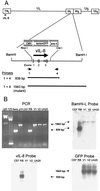
Structural characterization of RB1BvIL-8ΔsmGFP. (A) Schematic diagram of the MDV genome showing the region expressing the vIL-8 gene, the restriction sites used in the deletion of vIL-8, the smGFP expression cassette used in mutagenesis, and the location of PCR primers used in the structural characterization of the parental and mutant viruses. (B) An agarose gel showing the results of PCR amplification using primers 1 and 4 (shown in panel A) (upper left panel) and Southern blot hybridization analysis of these products using the BamHI-L fragment of the MDV genome (upper right panel), the vIL-8 cDNA (lower left panel), and the smGFP gene (lower right panel) as probes. The lanes are λd3, HindIII-digested lambda DNA molecular weight markers; 123, 123-bp DNA ladder markers (GIBCO-BRL); BamL, pUC19 containing the BamHI-L fragment of the MDV genome (20); pYH, transfer vector pYH6a; CEF, uninfected CEF DNA; RB, RB1B parental MDV-infected CEF DNA; V1, RB1BvIL-8ΔsmGFP clone 2-infected CEF DNA; V2, RB1BvIL-8ΔsmGFP reisolated from spleen cells of infected chickens; and UA20, DNA from cell line MDCC-UA20, established from an RB1BvIL-8ΔsmGFP-induced ovarian lymphoma.
FIG. 8
FIG. 8
Expression analysis of uninfected and RB1B- and RB1BvIL-8ΔsmGFP-infected CEF RNAs. The expression of cellular and virus gene products was examined using RT-PCR and primers specific for chicken β-actin (exon IV→exon V) (A); the smGFP gene (B); the MDV ICP27 gene (C); and the MDV vIL-8 gene (exon I→exon III) (D). Each picture shows the ethidium bromide-stained 1% agarose gel of 10 μl of each reaction product (total reaction volume, 50 μl). The templates used were lanes 1 to 3, 1 μg, 100 ng, and 10 ng of CEF total RNA, respectively; lanes 4 to 6, 1 μg, 100 ng, and 10 ng of RB1B-infected CEF total RNA, respectively; lanes 7 to 9, 1 μg, 100 ng, and 10 ng of RB1BvIL-8ΔsmGFP-infected CEF total RNA, respectively. Lane 10 in panel D represents 20 ng of RB1B-infected CEF DNA as a control for DNA contamination. HL denotes DNA Hyper-Ladder molecular size standard (Bioline). Sizes of specific RT-PCR products and molecular size standards are identified with arrows.
FIG. 9
FIG. 9
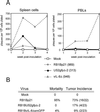
Comparison of RB1B and RB1BvIL-8ΔsmGFP, in vivo experiment 1. (A) Graphs of virus reisolation data for spleen cells and PBL from chickens (B15 × 7) inoculated with mock-infected CEF (Mock); equal-passage-level parent virus-infected CEF (RB1Bp21); RB1BUS2gfpΔ-2-infected CEF (US2gfpΔ-2); and RB1BvIL-8ΔsmGFP-infected CEF (vIL-8Δ). The numbers in parentheses indicate PFU inoculated per chicken. Each time point represents the mean plaque number counted on triplicate 60-mm-diameter dishes of CEF cocultivated with spleen cells or PBL at 6 days postplating. The y axis denotes plaques formed on CEF monolayers per 106 cell plated. (B) Summary of virus-specific mortality and tumor incidence data for this trial. Numbers in parentheses indicate the actual number of birds containing tumors divided by the number of birds left in treatment groups after virus reisolation attempts (i.e., after 4 weeks postinoculation).
FIG. 10
FIG. 10
Comparison of RB1B and RB1BvIL-8ΔsmGFP, in vivo experiment 2. (A) Graphic representations of virus reisolation data for spleen cells and PBL from commercial SPF chickens inoculated with mock-infected CEF (Mock); parent virus (RB1B)-infected CEF (RB1Bp12); and RB1BvIL-8ΔsmGFP-infected CEF (vIL-8Δ). Each time point represents the mean plaque number counted on triplicate 60-mm-diameter dishes of CEF cocultivated with spleen cells or PBL at 6 days postplating. The numbers in parentheses indicate PFU inoculated per chicken. The y axis denote plaques formed on CEF monolayers per 106 cell plated. (B) Summary of virus-specific mortality and tumor incidence data for this trial. Numbers in parentheses indicate the actual number of birds containing tumors divided by the number of birds left in treatment groups after virus reisolation attempts (i.e., after 4 weeks postinoculation).
FIG. 11
FIG. 11
Spontaneous expression of smGFP in MDCC-UA20 cells. Histogram plots of flow cytometry data are shown depicting the number and fluorescence intensity of GFP-expressing lymphoblastoid cells. The x axis denotes arbitrary fluorescence units (log scale) for the FL-1 (fluorescein isothiocyanate [FITC], GFP) channel, and the y axis denotes cell number. For each plot, 104 cells were acquired. The cell lines used were RECC-CU91, a REV-transformed T-lymphoblastoid cell line (53); MDCC-UA01, an RB1B-transformed T-lymphoblastoid cell line (18); MDCC-UA04, an RB1BUS2gfpΔ-1-transformed T-lymphoblastoid cell line (18); and MDCC-UA20, an RB1BvIL-8ΔsmGFP-transformed T-lymphoblastoid cell line.

Similar articles

Cited by

References

    1. Adams D H, Lloyd A R. Chemokines: leucocyte recruitment and activation cytokines. Lancet. 1997;349:490–495. - PubMed
    1. Adams D H, Shaw S. Leucocyte-endothelial interactions and regulation of leucocyte migration. Lancet. 1994;343:831–836. - PubMed
    1. Akiyama Y, Kato S, Iwa N. Continuous cell culture from lymphoma of Marek's disease. Biken J. 1973;16:177–179. - PubMed
    1. Ausubel F M, Brent R, Kingston R E, Moore D D, Seidman J G, Smith J A, Struhl K. Current protocols in molecular biology. New York, N.Y: John Wiley & Sons; 1995.
    1. Baggiolini M. Chemokines and leukocyte traffic. Nature. 1998;392:565–568. - PubMed

Publication types

MeSH terms

Associated data