

doi: 10.1128/JVI.75.11.5182-5188.2001.
Identification of the regions of Fv1 necessary for murine leukemia virus restriction
Affiliations
- PMID: 11333899
- PMCID: PMC114923
- DOI: 10.1128/JVI.75.11.5182-5188.2001
Item in Clipboard
Identification of the regions of Fv1 necessary for murine leukemia virus restriction
J Virol.
2001 Jun.
Abstract
The Fv1 gene restricts murine leukemia virus replication via an interaction with the viral capsid protein. To study this interaction, a number of mutations, including a series of N-terminal and C-terminal deletions, internal deletions, and a number of single-amino-acid substitutions, were introduced into the n and b alleles of the Fv1 gene and the effects of these changes on virus restriction were measured. A significant fraction of the Fv1 protein was not required for restriction; however, retention of an intact major homology region as well as of domains toward the N and C termini was essential. Binding specificity appeared to be a combinatorial property of a number of residues within the C-terminal portion of Fv1.
Figures
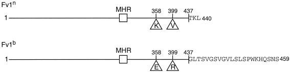
The differences between Fv1n and Fv1b proteins. The lines are to scale and represent the Fv1 gene product. The three positions that differ between Fv1n and Fv1b, residues 358 and 399 and the C terminus, are highlighted, with the amino acids in each case indicated by their single-letter codes. Sequences for Fv1n and Fv1b can be found under GenBank accession no. X97720 and no. X97719, respectively.
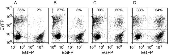
Fv1 activity of C-terminal deletion mutants. FACS profiles illustrating the effect of the C terminus on restriction of B-tropic tester virus are shown. EGFP expression is shown on the x axis; EYFP expression is shown on the y axis. The percentages given each indicate the proportion of EYFP-positive, i.e., B-MLV-infected, cells in the EGFP-negative (Fv1−) and the EGFP-positive (Fv1+) subpopulation. Wild-type Fv1n (A), mutant Fv1bΔ441–459 (B), wild-type Fv1b (C), and mutant Fv1bΔ411–459 (D) were introduced. The amount of restriction decreased from the full restriction shown in panel A to the partial restriction shown in panel B the slight restriction shown in panel C, and the lack of restriction shown in panel D. The percentages given are typical of the values for each level of restriction.
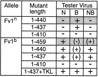
Summary of restriction by Fv1 C-terminal deletion mutants. The activity of each mutant against N-, B-, or NB-tropic tester virus is shown. The mutant length indicates at which residue the mutant terminates. The data for wild-type alleles are shaded. The extent of restriction is described by symbols, as follows: +, full restriction (equivalent to a reduction from 35% infection of Fv1− cells to 2% infection of Fv1+ cells, as shown by Fv1n restriction of B-MLV and Fv1b restriction of N-tropic MLV); (+), partial restriction; (−), slight restriction; −, no restriction.
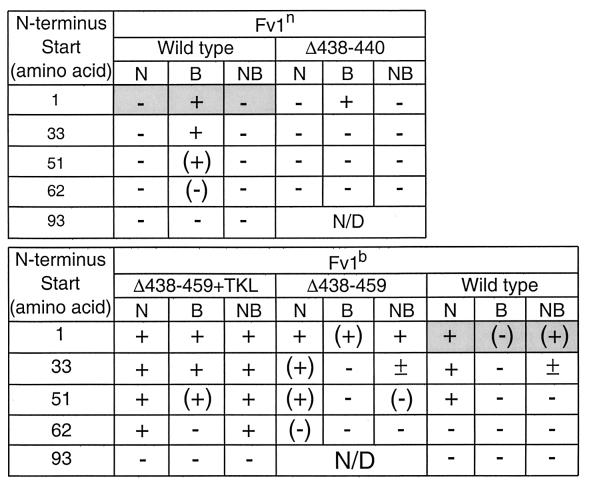
Restriction by Fv1 N-terminal deletion mutants. The activity of each mutant against N-, B-, or NB-tropic tester virus is shown. The data for wild-type alleles are shaded. The extent of restriction is indicated by symbols, as described in the legend to Fig. 3. ±, 50% restriction (half the percentage of Fv1+ cells are infected composed to the percentage infected in the Fv1− population).
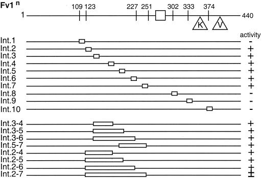
Restriction of Fv1n internal deletions. The diagram at the top shows the major features of Fv1n, with the lines below representing mutants. The stretch of sequence deleted is indicated (open boxes). The name of each mutant is shown to the left of each line, and its activity against B-tropic MLV is shown to the right. Symbols indicate the extent of restriction, as follows: +, complete; ±, 50% restriction −, none.

Restriction of Fv1 MHR mutants. The diagram shows the MHR sequence of wild-type Fv1 with the single-amino-acid changes made shown below. The consensus sequence is written at the bottom (Φ and O represent aromatic and aliphatic amino acids, respectively). The extent of restriction of Fv1n and Fv1b with each mutation is shown on the right. +, restriction; −, no restriction; n/d, not determined.
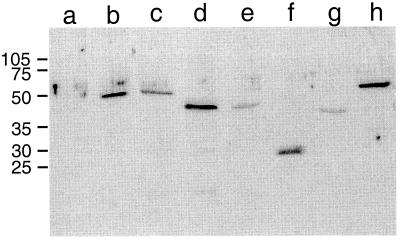
Detection of mutant Fv1 proteins. Shown are the results of Western blot analysis of M. dunni cells transduced with Fv1 derivatives Lanes: a, negative control; b, wild-type Fv1n; c, wild-type Fv1b; d, C-terminal deletion mutant Fv1nΔ1–410; e, C-terminal deletion mutant Fv1bΔ1–410; f, N-terminal deletion mutant Fv1nΔ1–201; g, internal deletion mutant Int.3–6; h, MHR mutant Fv1nE273D. Virus input was adjusted to result in 50 to 80% transduction levels as measured by EGFP expression. Total protein (100μg) was loaded in each lane, and Fv1 was detected with anti-Fv1 polyclonal antibody. Wild-type Fv1 and mutant Int. 3–6 are active; the remaining mutants are inactive.

Restriction of specificity mutants. Mutants are named with three letters; the first refers to position 358, the second to position 399, and the third to the C-terminus of the allele. n indicates the mutant is Fv1n-like at that position, b indicates it is Fv1b like at that position, A means that the residue has been mutated to alanine, and a dash in the third position indicates that the allele terminates at residue 437. Symbols are used to indicate the extent of restriction, as follows: +, full restriction; (+), partial restriction; (−), slight restriction; −, no restriction.
References
-
- Best S, Le Tissier P, Towers G, Stoye J P. Positional cloning of the mouse retrovirus restriction gene Fv1. Nature. 1996;382:826–829. - PubMed
-
- Campos-Olivas R, Newman J L, Summers M F. Solution structure and dynamics of the Rous Sarcoma Virus capsid protein and comparison with capsid proteins of other retroviruses. J Mol Biol. 2000;296:633–649. - PubMed
Publication types
MeSH terms
Substances
LinkOut - more resources
Full Text Sources
Other Literature Sources
