

doi: 10.1128/JVI.75.11.5370-5374.2001.
Active and selective transcytosis of cell-free human immunodeficiency virus through a tight polarized monolayer of human endometrial cells
Affiliations
- PMID: 11333919
- PMCID: PMC114943
- DOI: 10.1128/JVI.75.11.5370-5374.2001
Item in Clipboard
Active and selective transcytosis of cell-free human immunodeficiency virus through a tight polarized monolayer of human endometrial cells
J Virol.
2001 Jun.
Abstract
We report that both primary and laboratory-adapted infectious human immunodeficiency virus type 1 (HIV-1) isolates in a cell-free form are capable of transcytosis through a tight and polarized monolayer of human endometrial cells. Trancytosis of cell-free HIV occurs in a strain-selective fashion and appears to be dependent on interactions between HIV envelope glycoproteins and lectins on the apical membrane of the epithelial cells. These findings provide new insights into the initial events occurring during heterosexual transmission of the virus.
Figures
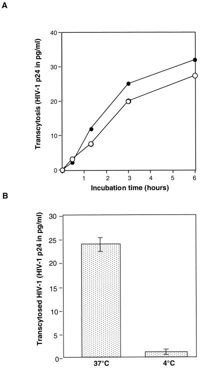
Transcytosis of cell-free and cell-associated HIV-1 through a tight monolayer of HEC-1 cells. (A) Kinetics of transcytosis of cell-free (full circles) and PBL-associated (open circles) HIV-1NDK. Twenty nanograms of p24 (free virus) and 2 × 10⁶ infected PBL were deposited in the apical chamber of the transwell system. The results are expressed as the amount of p24 antigen recovered in the basolateral chamber as a function of time. (B) Temperature dependency of transcytosis. Transcytosis of free HIV-1NDK through the HEC-1 cells monolayer was assessed at 37 and at 4°C by measuring the amount of p24 antigen in the basal chamber after 3 h of contact of cell-free virus (20 ng) with the apical membrane of HEC-1 cells. Results are expressed as means and standard deviations of three separate experiments.
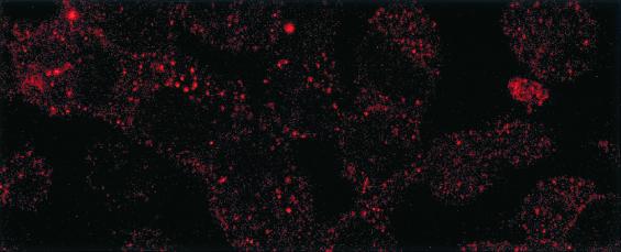
Detection of intracellular HIV-1 gp160 antigen (red) in transcytosed HEC-1 cells by immunoflorescence. The HEC-1 cells used in the transcytosis assays were washed, fixed with paraformaldehyde (4% in phosphate-buffered saline [PBS]) for 15 min, quenched of free aldehydes with 200 mM NH4Cl in PBS, and permeabilized for 10 min with 0.5% of Triton X-100 in PBS. After being washed with PBS, cells were incubated for 1 h with human anti-gp160 IgG diluted in PBS buffer with 1% bovine serum albumin. Phycoerythrin-labeled F(ab′)2 goat anti-human IgG (Jackson Immunoresearch, West Grove, Pa.) was further added at a dilution of 1/10. The coverslips were mounted in Mowiol (Sigma, St. Louis, Mo.) and observed by confocal microscopy using a Leica microscope (Leica, Wetzlar, Germany). Magnification, ×630.
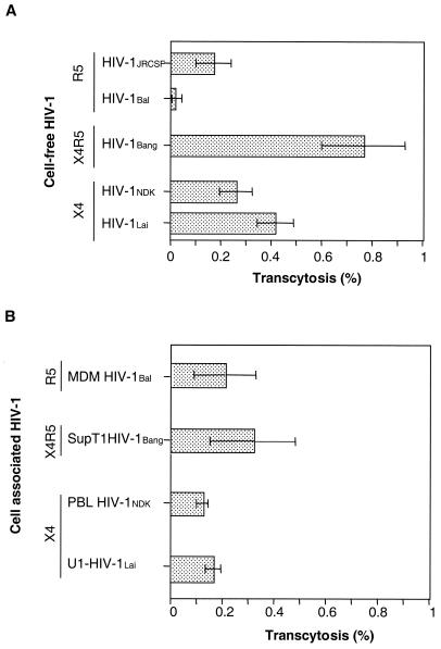
Transcytosis of various isolates of HIV-1 through HEC-1 cells. (A) Transcytosis of cell-free HIV. (B) Transcytosis of cell-associated HIV. The viral strains that were used included the primary R5-tropic HIV-1JRCSF (clade B) grown on PBL following stimulation with PHA and IL-2, the R5-tropic HIV-1Bal (clade B) which was amplified in monocyte-derived macrophages of healthy donors, the laboratory-adapted R5X4-tropic HIV-1Bang originating from a patient infected with clade A virus and further amplified in the Sup T1 T-cell line, the primary X4-tropic HIV-1NDK (clade D) grown in PBL of healthy donors following stimulation with PHA and IL-2, and the laboratory-adapted X4-tropic HIV-1Lai (clade B) amplified in U1 monocytic cells. Transcytosis was assessed as previously described (14). Filters were used when the resistivity of the monolayer had reached 200 Ω/cm2 after 6 days. Free virus (20 ng/well) or HIV-1-infected cells (2 × 10⁶ cells) were added to the apical chamber of transwells. After 180 min at 37°C, transcytosis was quantified by measuring the p24 antigen in samples taken from the basolateral chamber by means of a capture enzyme-linked immunosorbent assay (DuPont de Nemours, Wilmington, Del.) (threshold of detection, 3 pg/ml). Results were expressed as a percentage of virus recovered in the basal chamber, calculated from the amount of HIV-1 applied in the apical chamber that represented 100%. The data are expressed as means and standard deviations for three independent experiments.
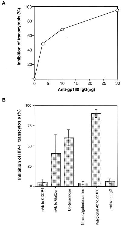
Inhibition of transcytosis of free HIV-1NDK through a tight HEC-1 epithelial barrier by anti-env and antireceptor antibodies. (A) Virus was incubated with serial amounts of purified polyclonal human antibodies to gp160 for 15 min at 37°C prior to being deposited in the apical chamber of the transwell system. (B) Cells were preincubated with 12G5 MAbs to CXCR4 (R & D Systems, Minneapolis, Minn.), and virus was incubated with rabbit polyclonal anti-Galcer antibodies (Sigma), d -(+)-mannose (Sigma), and N-acetylgalactosamine (Sigma). A positive control in the experiment was polyclonal IgG against gp160 purified from serum of HIV-1-seropositive individuals. The negative control was an irrelevant IgG purified from pooled serum of HIV-1-seronegative blood donors. The results are expressed as the percent inhibition of transcytosis and expressed as means ± standard deviations for four separate experiments.
Similar articles
-
Neutralizing monoclonal antibodies to human immunodeficiency virus type 1 do not inhibit viral transcytosis through mucosal epithelial cells.Virology. 2008 Jan 20;370(2):246-54. doi: 10.1016/j.virol.2007.09.006. Epub 2007 Oct 24. Virology. 2008. PMID: 17920650
-
R5- and X4-HIV-1 use differentially the endometrial epithelial cells HEC-1A to ensure their own spread: implication for mechanisms of sexual transmission.Virology. 2007 Feb 5;358(1):55-68. doi: 10.1016/j.virol.2006.07.029. Epub 2006 Aug 24. Virology. 2007. PMID: 16934308
-
Cell-to-cell contact results in a selective translocation of maternal human immunodeficiency virus type 1 quasispecies across a trophoblastic barrier by both transcytosis and infection.J Virol. 2001 May;75(10):4780-91. doi: 10.1128/JVI.75.10.4780-4791.2001. J Virol. 2001. PMID: 11312350 Free PMC article.
-
Infectious human immunodeficiency virus can rapidly penetrate a tight human epithelial barrier by transcytosis in a process impaired by mucosal immunoglobulins.J Infect Dis. 1999 May;179 Suppl 3:S448-53. doi: 10.1086/314802. J Infect Dis. 1999. PMID: 10099117 Review.
-
HIV-1 infection in the female reproductive tract: role of interactions between HIV-1 and genital epithelial cells.Am J Reprod Immunol. 2011 Mar;65(3):253-60. doi: 10.1111/j.1600-0897.2010.00965.x. Epub 2011 Jan 12. Am J Reprod Immunol. 2011. PMID: 21223427 Review.
Cited by
-
HSV-2- and HIV-1- permissive cell lines co-infected by HSV-2 and HIV-1 co-replicate HSV-2 and HIV-1 without production of HSV-2/HIV-1 pseudotype particles.Virol J. 2007 Jan 5;4:2. doi: 10.1186/1743-422X-4-2. Virol J. 2007. PMID: 17207276 Free PMC article.
-
Mother-to-Child HIV-1 Transmission Events Are Differentially Impacted by Breast Milk and Its Components from HIV-1-Infected Women.PLoS One. 2015 Dec 17;10(12):e0145150. doi: 10.1371/journal.pone.0145150. eCollection 2015. PLoS One. 2015. PMID: 26680219 Free PMC article.
-
Origin of the transmitted virus in HIV infection: infected cells versus cell-free virus.J Infect Dis. 2014 Dec 15;210 Suppl 3(Suppl 3):S667-73. doi: 10.1093/infdis/jiu369. J Infect Dis. 2014. PMID: 25414422 Free PMC article. Review.
-
The importance of semen leukocytes in HIV-1 transmission and the development of prevention strategies.Hum Vaccin Immunother. 2020 Sep 1;16(9):2018-2032. doi: 10.1080/21645515.2020.1765622. Epub 2020 Jul 2. Hum Vaccin Immunother. 2020. PMID: 32614649 Free PMC article. Review.
-
Adaptive HIV-specific B cell-derived humoral immune defenses of the intestinal mucosa in children exposed to HIV via breast-feeding.PLoS One. 2013 May 21;8(5):e63408. doi: 10.1371/journal.pone.0063408. Print 2013. PLoS One. 2013. PMID: 23704905 Free PMC article.
References
-
- Agace W W, Amara A, Roberts A I, Pablos J L, Thelen S, Uguccioni M, Li X Y, Marsal J, Arenzana-Seisdedos F, Delaunay T, Ebert E C, Moser B, Parker C M. Constitutive expression of stromal derived factor-1 by mucosal epithelia and its role in HIV transmission and propagation. Curr Biol. 2000;10:325–328. - PubMed
-
- Bomsel M. Transcytosis of infectious human immunodeficiency virus across a tight human epithelial cell line barrier. Nat Med. 1997;3:42–47. - PubMed
-
- Chenine A L, Matouskova E, Sanchez G, Reischig J, Pavlikova L, LeContel C, Chermann J C, Hirsch I. Primary intestinal epithelial cells can be infected with laboratory-adapted strain HIV type 1 NDK but not with clinical primary isolates. AIDS Res Hum Retrovir. 1998;14:1235–1238. - PubMed
-
- Collins K B, Patterson B K, Naus G J, Landers D V, Gupta P. Development of an in vitro organ culture model to study transmission of HIV-1 in the female genital tract. Nat Med. 2000;6:475–479. - PubMed
-
- Cook D G, Fantini J, Spitalnik S L, Gonzalez S F. Binding of human immunodeficiency virus type I (HIV-1) gp120 to galactosylceramide (GalCer): relationship to the V3 loop. Virology. 1994;201:206–214. - PubMed
Publication types
MeSH terms
Substances
LinkOut - more resources
Full Text Sources
Medical
