

Structural analysis of a fiber-pseudotyped adenovirus with ocular tropism suggests differential modes of cell receptor interactions
- PMID: 11333920
- PMCID: PMC114944
- DOI: 10.1128/JVI.75.11.5375-5380.2001
Structural analysis of a fiber-pseudotyped adenovirus with ocular tropism suggests differential modes of cell receptor interactions
Abstract
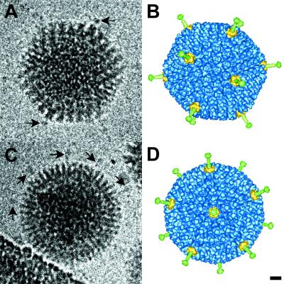
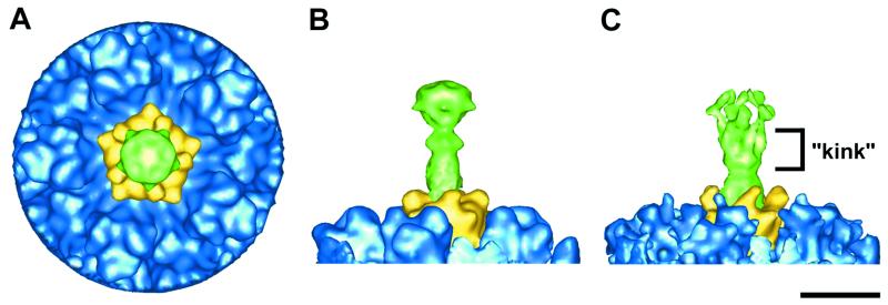
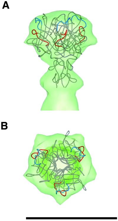
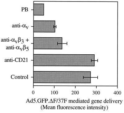
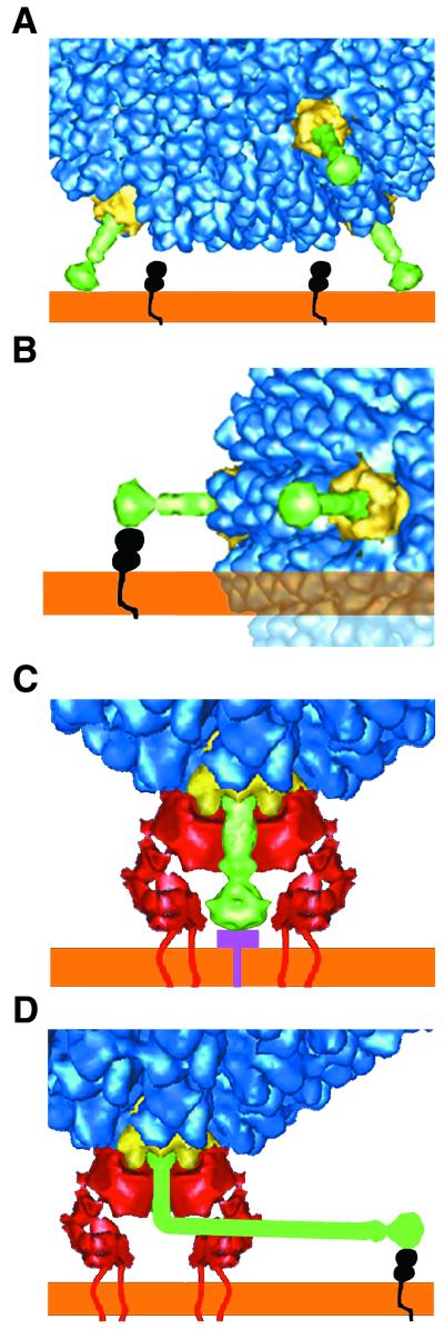
Adenovirus (Ad) entry into cells is initiated by the binding of the fiber knob to a cell surface receptor. The coxsackie- and adenovirus receptor (CAR) functions as the attachment receptor for many, but not all, Ad serotypes. Ad type 37 (Ad37), a subgroup D virus that causes keratoconjunctivitis in humans, does not infect cells via CAR despite demonstrated binding of the Ad37 knob to CAR. We have pseudotyped a fiber deletion Ad5 vector with the Ad37 fiber (Ad37f), and this vector retains the ocular tropism of Ad37. Here we present a cryo-electron microscopy reconstruction of Ad37f that shows the entire Ad37 fiber, including the shaft and knob domains. We have previously proposed that Ad37 may not utilize CAR for cell entry because of the geometric constraints imposed by a rigid fiber (E. Wu, J. Fernandez, S. K. Fleck, D. Von Seggern, S. Huang, and G. R. Nemerow, Virology 279:78-89, 2001). Consistent with this hypothesis, our structural results show that the Ad37 fiber is straight and rigid. Modeling of the interaction between Ad37f and host cell receptors indicates that fiber flexibility or rigidity, as well as length, can affect receptor usage and cellular tropism.
Figures
Similar articles
-
Flexibility of the adenovirus fiber is required for efficient receptor interaction.J Virol. 2003 Jul;77(13):7225-35. doi: 10.1128/jvi.77.13.7225-7235.2003. J Virol. 2003. PMID: 12805421 Free PMC article.
-
Dependence of adenovirus infectivity on length of the fiber shaft domain.J Virol. 2000 Nov;74(22):10274-86. doi: 10.1128/jvi.74.22.10274-10286.2000. J Virol. 2000. PMID: 11044071 Free PMC article.
-
Adenovirus type 9 fiber knob binds to the coxsackie B virus-adenovirus receptor (CAR) with lower affinity than fiber knobs of other CAR-binding adenovirus serotypes.J Virol. 2001 Aug;75(15):7210-4. doi: 10.1128/JVI.75.15.7210-7214.2001. J Virol. 2001. PMID: 11435605 Free PMC article.
-
Tropism and transduction of oncolytic adenovirus 5 vectors in cancer therapy: Focus on fiber chimerism and mosaicism, hexon and pIX.Virus Res. 2018 Sep 15;257:40-51. doi: 10.1016/j.virusres.2018.08.012. Epub 2018 Aug 17. Virus Res. 2018. PMID: 30125593 Review.
-
Adenovirus interaction with its cellular receptor CAR.Curr Top Microbiol Immunol. 2003;272:331-64. doi: 10.1007/978-3-662-05597-7_11. Curr Top Microbiol Immunol. 2003. PMID: 12747555 Review.
Cited by
-
Model of the trimeric fiber and its interactions with the pentameric penton base of human adenovirus by cryo-electron microscopy.J Mol Biol. 2011 Mar 11;406(5):764-74. doi: 10.1016/j.jmb.2010.11.043. Epub 2010 Dec 10. J Mol Biol. 2011. PMID: 21146538 Free PMC article.
-
Adenovirus.Curr Top Microbiol Immunol. 2010;343:195-224. doi: 10.1007/82_2010_16. Curr Top Microbiol Immunol. 2010. PMID: 20376613 Free PMC article. Review.
-
Flexibility of the adenovirus fiber is required for efficient receptor interaction.J Virol. 2003 Jul;77(13):7225-35. doi: 10.1128/jvi.77.13.7225-7235.2003. J Virol. 2003. PMID: 12805421 Free PMC article.
-
Selection Pressure in the Human Adenovirus Fiber Knob Drives Cell Specificity in Epidemic Keratoconjunctivitis.J Virol. 2016 Oct 14;90(21):9598-9607. doi: 10.1128/JVI.01010-16. Print 2016 Nov 1. J Virol. 2016. PMID: 27512073 Free PMC article.
-
The cell adhesion molecule "CAR" and sialic acid on human erythrocytes influence adenovirus in vivo biodistribution.PLoS Pathog. 2009 Jan;5(1):e1000277. doi: 10.1371/journal.ppat.1000277. Epub 2009 Jan 2. PLoS Pathog. 2009. PMID: 19119424 Free PMC article.
References
-
- Adrian M, Dubochet J, Lepault J, McDowall A W. Cryo-electron microscopy of viruses. Nature. 1984;308:32–36. - PubMed
-
- Arnberg N, Mei Y, Wadell G. Fiber genes of adenoviruses with tropism for the eye and the genital tract. Virology. 1997;227:239–244. - PubMed
-
- Bergelson J M, Cunningham J A, Droguett G, Kurt-Jones E A, Krithivas A, Hong J S, Horwitz M S, Crowell R L, Finberg R W. Isolation of a common receptor for coxsackie B viruses and adenoviruses 2 and 5. Science. 1997;275:1320–1323. - PubMed
Publication types
MeSH terms
Substances
Grants and funding
LinkOut - more resources
Full Text Sources
Other Literature Sources
