

Review
Multi-slice proton MR spectroscopy and diffusion-weighted imaging in methylmalonic acidemia: report of two cases and review of the literature
Affiliations
- PMID: 11337323
- PMCID: PMC8174958
Item in Clipboard
Review
Multi-slice proton MR spectroscopy and diffusion-weighted imaging in methylmalonic acidemia: report of two cases and review of the literature
AJNR Am J Neuroradiol.
2001 May.
Abstract
Methylmalonic acidemia is an inborn disorder of amino acid metabolism that commonly presents with neurologic deficits. We present the results of multi-slice proton MR spectroscopy and diffusion-weighted imaging of the brain in two patients with methylmalonic acidemia. The findings consisted of restricted diffusion and elevated lactate in the globi pallidi, compatible with acute infarction (patient 1) and elevated lactate in CSF (patient 2).
Figures
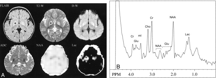
Patient 1. A, Symmetrical high signal intensity in the globi pallidi is demonstrated (*) on fast-FLAIR (8800/140/2200 [TR/TEeff/TI]) and T2-weighted FSE (5015/97 [TR/TEeff]) MR images. On the diffusion-weighted (10,000/100 [TR/TE]) (b = 1000 s/mm2) MR image, the lesions were mildly hyperintense anteromedially and markedly hyperintense posterolaterally (arrowheads). The ADCave map demonstrated symmetrical low signal intensity (restricted diffusion) in the posterolateral portions of the globi pallidi (arrowheads), consistent with acute infarctions. Metabolic images showed symmetrical bilateral decreases in NAA and increases in Lac in globi pallidi (*), also consistent with acute infarction. Other brain regions appeared normal, and no Lac was detected in CSF. B, Single-voxel short-TE spectrum (2000/30 [TR/TE]) of the left globus pallidus shows decreased levels of NAA and increased levels of Lac compared with controls (not shown). Levels of other metabolites are within normal limits (Glu, glutamate and glutamine; mI, myo-inositol)
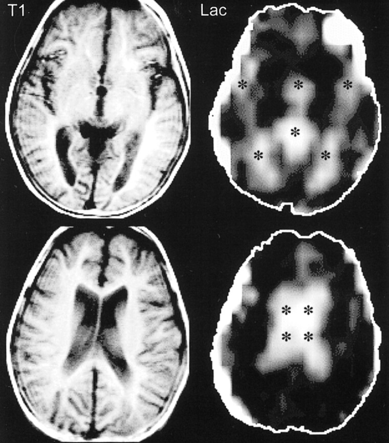
Patient 2. T1-weighted spin-echo MR images (300/13 [TR/TE]) at the level of the third ventricle (top row) and the lateral ventricles (bottom row) were unremarkable except for mild volume loss. Lac images showed high signal limited to the CSF spaces (*) (third and lateral ventricles, sylvian fissures, and cistern of the velum interpositum)
References
-
- van der Meer SB, Poggi F, Spada M, et al. Clinical outcome of long-term management of patients with vitamin B12 unresponsive methylmalonic acidemia. J Pediatr 1994;125:903-908 - PubMed
-
- Lam WW, Wang ZJ, Zhao H, et al. 1H MR spectroscopy of the basal ganglia in childhood: a semiquantitative analysis. Neuroradiology 1998;40:315-323 - PubMed
-
- Wajner M, Coelho JC. Neurological dysfunction in methylmalonic acidaemia is probably related to the inhibitory effect of methylmalonate on brain energy production. J Inherit Metab Dis 1997;20:761-768 - PubMed
Publication types
MeSH terms
Substances
LinkOut - more resources
Full Text Sources
Medical