Combined benign odontogenic tumors: CT and MR findings and histomorphologic evaluation
- PMID: 11337330
- PMCID: PMC8174925
Combined benign odontogenic tumors: CT and MR findings and histomorphologic evaluation
Abstract
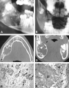
Calcifying epithelial odontogenic tumors and calcifying odontogenic cysts are rare, benign odontogenic tumors. We report two cases of an exceptional combination of these tumors with either an ameloblastic fibroodontoma or an odontoma.
Figures


Similar articles
-
[Mixed odontogenic tumours].Rev Stomatol Chir Maxillofac. 2009 Sep;110(4):217-20. doi: 10.1016/j.stomax.2009.06.005. Epub 2009 Aug 5. Rev Stomatol Chir Maxillofac. 2009. PMID: 19660774 French.
-
[Mixed odontogenic tumors in children and adolescents].Fogorv Sz. 2007 Apr;100(2):65-9. Fogorv Sz. 2007. PMID: 17546897 Hungarian.
-
Ameloblastic fibro-odontoma masquerading as odontoma.Indian J Dent Res. 2011 Jul-Aug;22(4):616. doi: 10.4103/0970-9290.90332. Indian J Dent Res. 2011. PMID: 22124075
-
CT and MR imaging appearances of an extraosseous calcifying epithelial odontogenic tumor (Pindborg tumor).AJNR Am J Neuroradiol. 2000 Feb;21(2):343-5. AJNR Am J Neuroradiol. 2000. PMID: 10696021 Free PMC article. Review.
-
Calcifying odontogenic cyst associated with complex odontoma: case report and review of the literature.Med Oral Patol Oral Cir Bucal. 2005 May-Jul;10(3):243-7. Med Oral Patol Oral Cir Bucal. 2005. PMID: 15876968 Review. English, Spanish.
Cited by
-
Large erupted complex odontoma mimicking maxillary osteomyelitis.BMJ Case Rep. 2023 Jan 20;16(1):e253322. doi: 10.1136/bcr-2022-253322. BMJ Case Rep. 2023. PMID: 36669786 Free PMC article.
-
Anomalous morphology of an ectopic tooth in the maxillary sinus on three-dimensional computed tomography images.J Radiol Case Rep. 2013 Feb 1;7(2):11-6. doi: 10.3941/jrcr.v7i2.1227. Print 2013 Feb. J Radiol Case Rep. 2013. PMID: 23705035 Free PMC article.
-
[An irregular radio-opaque mass occupying the maxillary sinus. A specialist referral from the dentist].HNO. 2014 Apr;62(4):286-9. doi: 10.1007/s00106-013-2831-z. HNO. 2014. PMID: 24633382 German. No abstract available.
-
Calcifying odontogenic cyst with atypical features.Ann Maxillofac Surg. 2012 Jan;2(1):82-5. doi: 10.4103/2231-0746.95331. Ann Maxillofac Surg. 2012. PMID: 23482835 Free PMC article.
-
Mandibular hybrid odontogenic tumor.Oral Maxillofac Surg. 2025 Jan 24;29(1):45. doi: 10.1007/s10006-024-01329-9. Oral Maxillofac Surg. 2025. PMID: 39853419
References
-
- Pindborg JJ. Calcifying epithelial odontogenic tumors. Acta Pathol Microbiol Scand (Suppl) 1955;111:71 - PubMed
-
- Gorlin RJ, Pindborg JJ, Clausen FP, Vixkers RA. The calcifying odontogenic cyst: a possible analogue of the cutaneous epithelioma of Malherbe. Oral Surg 1962;15:1235-1243 - PubMed
-
- Pindborg JJ, Kramer IRH. International Histological Classification of Tumours: Histological Typing of Odontogenic Tumours, Jaw Cysts and Allied Lesions.. Geneva: World Health Organization, 1971
-
- Kramer RH, Pindborg JJ, Shear M. International Histological Classification of Tumours: Histological Typing of Odontogenic Tumours. 2nd ed. Berlin: Springer; 1992
-
- Kramer IRH, Pindborg JJ, Shear M. The WHO international histological classification of tumours: a commentary on the second edition. Cancer 1992;70:2988-2994 - PubMed
Publication types
MeSH terms
LinkOut - more resources
Full Text Sources
Medical
