

Diffusion-weighted MR imaging of intracerebral masses: comparison with conventional MR imaging and histologic findings
- PMID: 11337344
- PMCID: PMC8174938
Diffusion-weighted MR imaging of intracerebral masses: comparison with conventional MR imaging and histologic findings
Abstract
Background and purpose: The purposes of this study were to find the role of diffusion-weighted MR imaging in characterizing intracerebral masses and to find a correlation, if any, between the different parameters of diffusion-weighted imaging and histologic analysis of tumors. The usefulness of diffusion-weighted imaging and apparent diffusion coefficient (ADC) maps in tumor delineation was evaluated. Contrast with white matter and ADC values for tumor components with available histology were also evaluated.
Methods: Twenty patients with clinical and routine MR imaging/CT evidence of intracerebral neoplasm were examined with routine MR imaging and echo-planar diffusion-weighted imaging. The routine MR imaging included at least the axial T2-weighted fast spin-echo and axial T1-weighted spin-echo sequences before and after contrast enhancement. The diffusion-weighted imaging included an echo-planar spin-echo sequence with three b values (0, 300, and 1200 s/mm(2)), sensitizing gradient in the z direction, and calculated ADC maps. The visual comparison of routine MR images with diffusion-weighted images for tumor delineation was performed as was the statistical analysis of quantitative diffusion-weighted imaging parameters with histologic evaluation.
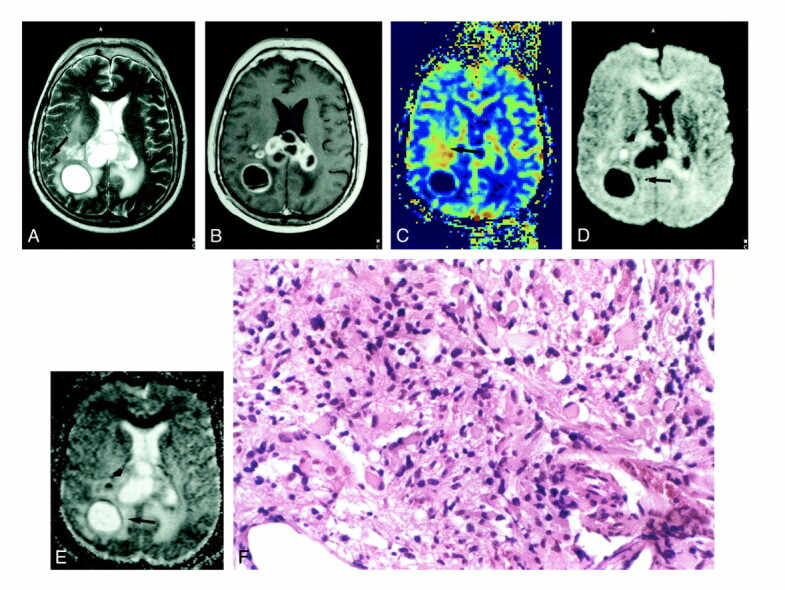
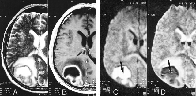
Results: For tumors, the diffusion-weighted images and ADC maps of gliomas were less useful than the T2-weighted spin-echo and contrast-enhanced T1-weighted spin-echo images in definition of tumor boundaries. Additionally, in six cases of gliomas, neither T2-weighted spin-echo nor diffusion-weighted images were able to show a boundary between tumor and edema, which was present on contrast-enhanced T1-weighted and/or perfusion echo-planar images. The ADC values of solid gliomas, metastases, and meningioma were in the same range. In two cases of lymphomas, there was a good contrast with white matter, with strongly reduced ADC values. For infection, the highest contrast on diffusion-weighted images and lowest ADC values were observed in association with inflammatory granuloma and abscess.
Conclusion: Contrary to the findings of previous studies, we found no clear advantage of diffusion-weighted echo-planar imaging in the evaluation of tumor extension. The contrast between gliomas, metastases, meningioma, and white matter was generally lower on diffusion-weighted images and ADC maps compared with conventional MR imaging. Unlike gliomas, the two cases of lymphomas showed hyperintense signal on diffusion-weighted images whereas the case of cerebral abscess showed the highest contrast on diffusion-weighted images with very low ADC values. Further study is required to find out whether this may be useful in the differentiation of gliomas and metastasis from lymphoma and abscess.
Figures


References
-
- Benveniste H, Hedlund LW, Johnson GA. Mechanism of detection of acute cerebral ischemia in rats by diffusion-weighted magnetic resonance microscopy. Stroke 1992;23:746-754 - PubMed
-
- Hossmann K-A, Fischer M, Bockhorst K, Hoehn-Berlage M. NMR imaging of the apparent diffusion coefficient (ADC) for the evaluation of metabolic suppression and recovery after prolonged cerebral ischemia. J Cereb Blood Flow Metab 1994;14:723-731 - PubMed
-
- Tien RD, Felsberg GJ, Friedman H, Brown M, MacFall J. MR imaging of high-grade cerebral gliomas: value of diffusion-weighted echoplanar pulse sequences. AJR Am J Roentgenol 1994;162:671-677 - PubMed
Publication types
MeSH terms
LinkOut - more resources
Full Text Sources
Medical