. 2001 May;22(5):977-84.

Initial and follow-up MR imaging findings in AIDS-related progressive multifocal leukoencephalopathy treated with highly active antiretroviral therapy

Affiliations

Initial and follow-up MR imaging findings in AIDS-related progressive multifocal leukoencephalopathy treated with highly active antiretroviral therapy

M M Thurnher et al. AJNR Am J Neuroradiol. 2001 May.

Abstract

Background and purpose: Recent studies have shown the beneficial effect of highly active antiretroviral therapy (HAART) in AIDS-related progressive multifocal leukoencephalopathy (PML). The purpose of our study was to evaluate the initial and follow-up imaging findings and survival in patients with PML who were treated with HAART.

Methods: The clinical course and MR imaging findings on initial and follow-up MR studies in four consecutive AIDS patients with PML who were treated with HAART are described.

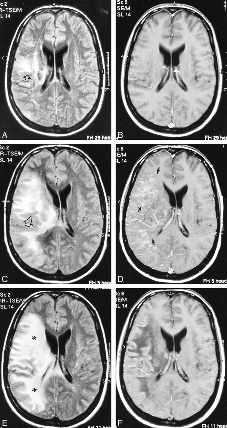
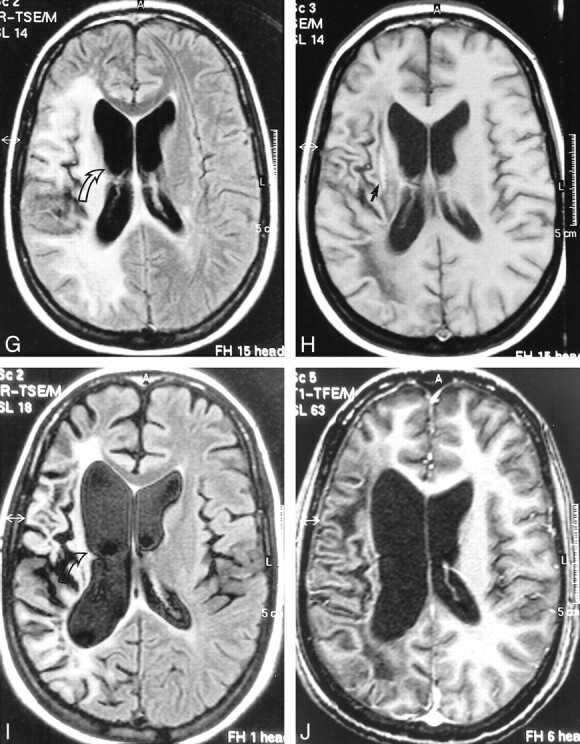
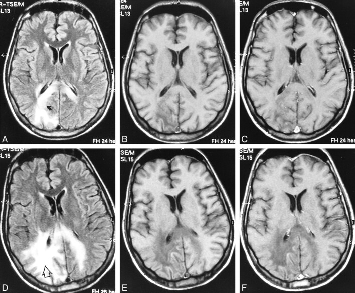
Results: Two patients were short-term survivors and died after 3 months. Two patients are still alive, with a survival time of 22 and 43 months, respectively. On initial MR studies, more extensive white matter changes were seen in the short-term survivors. Development of a mass effect and temporary enhancement (in one patient) was observed in two HAART responders on follow-up MR studies. Increased hypointensity on T1-weighted images with concomitant low signal on fluid-attenuated inversion-recovery fast spin-echo (FLAIR-FSE) images was seen in two responders, representing leukomalacia. Atrophic changes of the involved areas of the brain, consistent with burnt out PML lesions, were seen in two long-term survivors. In the short-term survivors, increased hypointensity was present on T1-weighted images with increased high signal on FLAIR-FSE images, representing progressive destructive disease.

Conclusion: Our results suggest that a clinical and radiologic response can be seen in some patients with AIDS-associated PML on HAART while in others there may be no beneficial response. Development of a mass effect and temporary enhancement on MR images in the early phase of treatment might represent positive predictive factors for prolonged survival.


Figures

<sc>fig</sc> 1.
fig 1.
Case 2: 32-year-old HIV-positive woman with PML and survival time exceeding 22 months. A and B, Initial MR study, December 1998. Axial FLAIR-FSE (TR/TE = 10000/150, TI = 2600) image (A) shows high-signal-intensity lesion in the white matter of the right centrum semiovale (arrow). There is no mass effect. No enhancement was present on contrast-enhanced T1-weighted (550/20) image (B). C and D, Follow-up MR study, January 1999 (1 month after initiation of HAART). FLAIR-FSE (10000/150, TI = 2600) image (C) shows progression of white matter abnormalities (arrow) with mass effect and compression of right ventricle. Increased hypoattenuation of the lesion was evident on T1-weighted (550/20) images (not shown). On contrast-enhanced T1-weighted (550/20) image (D), diffuse enhancement of white matter abnormalities is present (arrows). E and F, Subsequent MR study, February 1999 (2 months after initiation of HAART). FLAIR-FSE (10000/150, TI = 2600) image (E) shows further progression of the high-signal-intensity lesions of white matter disease (asterisks). Slight enhancement is still present on contrast-enhanced T1-weighted (550/20) image (F). Note increased hypoattenuation of the lesions and regression of the mass effect. Continued on page 982
<sc>fig</sc> 1.
fig 1.
Continued. G and H, Further follow-up MR study, April 1999 (4 months after initiation of HAART). FLAIR-FSE (10000/150, TI = 2600) image (G) reveals regression of the white matter changes with partially present low signal (corresponding to areas of increased hypoattenuation on T1-weighted images). Note atrophic changes of the right hemisphere with widening of the ventricle (arrow). Further increase in hypointensity is evident on T1-weighted (550/20) image (H). Elongated hyperintense area is seen medial to the sylvian fissure, representing subacute hemorrhage (arrow). Enhancement was not present on contrast-enhanced T1-weighted image (not shown). I and J, Follow-up MR examination, September 2000 (21 months after initiation of HAART). Axial FLAIR-FSE (7384/130, TI = 2100) image (I) and contrast-enhanced T1-weighted (8/2.7, flip angle = 10°) image (J) show atrophic changes in the right hemisphere with widening of the sulci and further ex-vacuo widening of the right ventricle (arrow). Note low signal on the FLAIR-FSE image, representing leukomalacia
<sc>fig</sc> 2.
fig 2.
Case 4: 28-year-old man with AIDS who presented with psychomotor slowing and hemianopsia. Stereotactic biopsy revealed PML. A–C, Initial MR study, March 1998. Axial FLAIR-FSE (10000/150, TI = 2600) image (A) shows scalloped, high-signal-intensity lesion in the parietooccipital white matter with extension to dorsal part of the corpus callosum on both sides (arrow). There is no mass effect. The lesion is hypointense on noncontrast T1-weighted SE (550/20) image (B). No enhancement is observed on contrast-enhanced T1-weighted SE (550/20) image (C). D–F, Follow-up MR study, May 1998 (4 weeks after the start of HAART). FLAIR-FSE (10000/150, TI = 2600) image (D) shows progression of the white matter lesion (arrow) with involvement of the right parietal white matter and contralateral occipital white matter. Increased hypointensity is evident on noncontrast T1-weighted (550/20) image (E). Contrast-enhanced T1-weighted (550/20) image shows no enhancement (F)

Similar articles

Cited by

References

    1. Berger JR, Concha M. Progressive multifocal leukoencephalopathy: the evolution of a disease once considered rare. Neurovirol 1995;1:5-18 - PubMed
    1. Happe S, Besselmann M, Matheja P, et al. Therapy of progressive multifocal leukoencephalopathy (PML) in AIDS with cidofovir (Vistide): review of the literature and two case reports. Nervenarzt 1999;70:935-943 - PubMed
    1. Portegies P, Algra PR, Hollak CEM, et al. Response to cytarabine in progressive multifocal leukoencephalopathy in AIDS. Lancet 1991;337:680-681 - PubMed
    1. Nicoli F, Chave B, Peragut JC, Gastaut JL. Efficacy of cytarabine in progressive multifocal leukoencephalopathy in AIDS. Lancet 1991;339:306 - PubMed
    1. De Luca A, Giancola ML, Cingolani A, et al. Clinical and virological monitoring during treatment with intrathecal cytarabine in patients with AIDS-associated progressive multifocal leukoencephalopathy. Clin Infect Dis 1999;28:624-628 - PubMed

MeSH terms