

Expression and localization of a novel Rab small G protein (Rab38) in the rat lung
- PMID: 11337364
- PMCID: PMC1891947
- DOI: 10.1016/S0002-9440(10)64122-3
Expression and localization of a novel Rab small G protein (Rab38) in the rat lung
Abstract
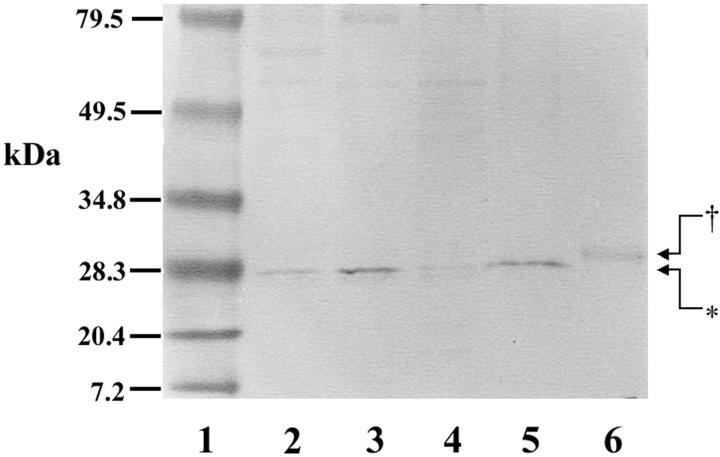
The Rab small G protein family participates in intracellular vesicle transport, including exocytosis and endocytosis. The cDNA encoding a novel Rab-related small G protein (Rab38) has been cloned from rat lung cDNA library and recorded in GenBank (accession no. M94043). However, the expression and localization of the protein in the lung remains primarily unknown. We produced polyhistidine-tagged recombinant Rab38 and a polyclonal antibody with a synthetic peptide. Immunohistochemistry demonstrated that the protein is specifically localized in alveolar type II cells and in bronchial epithelial cells. In situ hybridization using a digoxygenin-labeled RNA riboprobe clearly showed that the mRNA of the protein is localized in alveolar type II cells and bronchial epithelial cells, especially terminal airway epithelial cells. Western blot and reverse transcriptase-polymerase chain reaction showed distinct expression of the protein and mRNA in isolated alveolar type II cells, but not in alveolar macrophages. The native protein was predominantly hydrophobic and was enriched in a high-density vesicle fraction but was barely detectable in nuclear and lamellar body fractions in alveolar type II cells. Immunofluorescence cytochemistry performed on cultured alveolar type II cells showed that Rab38 distributed extensively in the cytoplasm with a distribution pattern similar to endoplasmic reticulum rather than other subcellular organelles. These results suggest that this novel rab small G protein (Rab38) mediates vesicular transport in terminal airway epithelium.
Figures


Similar articles
-
Exogenous gene transfer of Rab38 small GTPase ameliorates aberrant lung surfactant homeostasis in Ruby rats.Respir Res. 2017 Apr 24;18(1):70. doi: 10.1186/s12931-017-0549-2. Respir Res. 2017. PMID: 28438206 Free PMC article.
-
A mutation in Rab38 small GTPase causes abnormal lung surfactant homeostasis and aberrant alveolar structure in mice.Am J Pathol. 2008 Nov;173(5):1265-74. doi: 10.2353/ajpath.2008.080056. Epub 2008 Oct 2. Am J Pathol. 2008. PMID: 18832574 Free PMC article.
-
Expression and characterization of Rab38, a new member of the Rab small G protein family.Biol Chem. 2005 Feb;386(2):143-53. doi: 10.1515/BC.2005.018. Biol Chem. 2005. PMID: 15843158
-
Rab38 targets to lamellar bodies and normalizes their sizes in lung alveolar type II epithelial cells.Am J Physiol Lung Cell Mol Physiol. 2011 Oct;301(4):L461-77. doi: 10.1152/ajplung.00056.2011. Epub 2011 Jul 15. Am J Physiol Lung Cell Mol Physiol. 2011. PMID: 21764986 Free PMC article.
-
A plasmid library of full-length zebrafish rab proteins for in vivo cell biology.Cell Logist. 2017 Mar 1;7(1):e1301151. doi: 10.1080/21592799.2017.1301151. eCollection 2017. Cell Logist. 2017. PMID: 28396820 Free PMC article. Review.
Cited by
-
Exogenous gene transfer of Rab38 small GTPase ameliorates aberrant lung surfactant homeostasis in Ruby rats.Respir Res. 2017 Apr 24;18(1):70. doi: 10.1186/s12931-017-0549-2. Respir Res. 2017. PMID: 28438206 Free PMC article.
-
Rab38 and Rab32 control post-Golgi trafficking of melanogenic enzymes.J Cell Biol. 2006 Oct 23;175(2):271-81. doi: 10.1083/jcb.200606050. Epub 2006 Oct 16. J Cell Biol. 2006. PMID: 17043139 Free PMC article.
-
High expression of RAB38 promotes malignant progression of pancreatic cancer.Mol Med Rep. 2019 Feb;19(2):909-918. doi: 10.3892/mmr.2018.9732. Epub 2018 Dec 10. Mol Med Rep. 2019. PMID: 30569114 Free PMC article.
-
A mutation in Rab38 small GTPase causes abnormal lung surfactant homeostasis and aberrant alveolar structure in mice.Am J Pathol. 2008 Nov;173(5):1265-74. doi: 10.2353/ajpath.2008.080056. Epub 2008 Oct 2. Am J Pathol. 2008. PMID: 18832574 Free PMC article.
-
Darkness descends with two Rabs.J Cell Biol. 2006 Oct 23;175(2):199-200. doi: 10.1083/jcb.200608058. Epub 2006 Oct 16. J Cell Biol. 2006. PMID: 17043141 Free PMC article.
References
-
- Takai Y, Kaibuchi K, Kikuchi A, Kawata M: Small GTP-binding proteins. Int Rev Cytol 1992, 133:187-230 - PubMed
-
- Olkkonen VM, Stenmark H: Role of Rab GTPases in membrane traffic. Int Rev Cytol 1997, 176:1-85 - PubMed
-
- Novick P, Brennwald P: Friends and family: the role of the Rab GTPases in vesicular traffic. Cell 1993, 75:597-601 - PubMed
-
- Jager D, Stockert E, Jager E, Gure AO, Scanlan MJ, Knuth A, Old LJ, Chen YT: Serological cloning of a melanocyte rab guanosine 5′-triphosphate-binding protein and a chromosome condensation protein from a melanoma complementary DNA library. Cancer Res 2000, 60:3584-3591 - PubMed
-
- Dobbs LG, Geppert EF, Williams MC, Greenleaf RD, Mason RJ: Metabolic properties and ultrastructure of alveolar type II cells isolated with elastase. Biochim Biophys Acta 1980, 618:510-523 - PubMed
Publication types
MeSH terms
Substances
Associated data
- Actions
LinkOut - more resources
Full Text Sources
Molecular Biology Databases
