. 2001 May;158(5):1685-94.
doi: 10.1016/s0002-9440(10)64124-7.

Expression of a novel RNA-splicing factor, RA301/Tra2beta, in vascular lesions and its role in smooth muscle cell proliferation

Affiliations

Expression of a novel RNA-splicing factor, RA301/Tra2beta, in vascular lesions and its role in smooth muscle cell proliferation

Y Tsukamoto et al. Am J Pathol. 2001 May.

Abstract

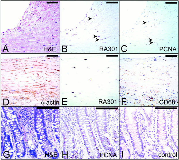
RA301/Tra2beta, a sequence-specific RNA-binding protein, was first cloned as a stress molecule in re-oxygenated astrocytes. In human vascular tissues, we have found enhanced RA301/Tra2beta expression in coronary artery with intimal thickening, and atherosclerotic aorta. Balloon injury to the rat carotid artery induced RA301/Tra2beta transcripts followed by expression of the antigen, which was detected in medial and neointimal vascular smooth muscle cells (VSMCs). In cultured VSMCs, hypoxia/re-oxygenation caused induction of RA301/Tra2beta and was accompanied by cell proliferation, both of which were blocked by the addition of either diphenyl iodonium, a NADPH oxidase inhibitor, PD98059, a mitogen-activated protein kinase kinase inhibitor, or antisense oligonucleotide for RA301/Tra2beta. Consistent with a link between RA301/Tra2beta and cell proliferation, platelet-derived growth factor also induced expression of RA301/Tra2beta in cultured VSMCS: These data suggest a possible role for RA301/Tra2beta in the regulation of VSMC proliferation, especially in the setting of hypoxia/re-oxygenation-induced cell stress.


Figures

Figure 1.
Figure 1.
Northern blotting for RA301/Tra2β mRNA in human arteries. Total RNA was extracted from human vascular tissues, subjected to electrophoresis (10 μg/lane) and transferred to nylon membranes. Northern blotting was performed using either a 32P-dCTP-labeled human RA301/Tra2β probe (top) or human β-actin probe (bottom). The numbers above lanes represent samples prepared from human spleen (lane 1, positive control), aorta from a 0-year-old male who died of congenital heart disease (lane 2), aorta of a 1-year-old male who died of congenital heart disease (lane 3), aorta of a 13-year-old male who died of congestive heart failure because of dilated cardiomyopathy (lane 4), aorta of a 31-year-old female who died of congestive heart failure because of cardiac sarcoma (lane 5), aorta of a 44-year-old male who died of congestive heart failure because of dilated cardiomyopathy (lane 6), aorta of a 55-year-old female who died of congestive heart failure because of dilated cardiomyopathy (lane 7), aorta from a 58-year-old male who died of cerebral hemorrhage because of hypertension and had a past history of hypercholesterolemia (lane 8), coronary artery from a 63-year-old male who died of gastric cancer and hepatocellular carcinoma, and had past history of hypertension and angina pectoris (lane 9), aorta of a 69-year-old male who died of cardioembolic brain infarction (lane 10), aorta of an 82-year-old male who died of congestive heart failure because of combined valvular disease (lane 11), and aorta of an 81-year-old female who died of cardioembolic brain infarction. The migration of ribosomal RNA is indicated on the left. Arrowheads indicate the splicing variants of RA301/RA301/Tra2β transcripts.
Figure 2.
Figure 2.
Expression of human RA301/Tra2β message in human coronary artery. A coronary artery was obtained from the same patient as described in Figure 1 ▶ , lane 9. A to C and D to F are adjacent sections, respectively. Sections of coronary artery were subjected to in situ hybridization using UTP-digoxigenin-labeled RA301/Tra2β riboprobes (B and E), and immunohistochemistry using anti-PCNA antibody (C, counterstained with hematoxylin), anti-α-smooth muscle actin antibody (1A4) (D) and anti-CD68 antibody (PG-M1) (F). A was shown as an H&E staining for orientation. Arrowheads in B and C are same cells positive for both RA301 message and PCNA antigen. RA301 mRNA-positive cells in E are not stained with anti-CD68 antibody but stained with anti-α-smooth muscle actin in adjacent sections (F and D). Sections of human colon from surgical specimen were immunostained with anti-PCNA antibody or preimmune serum, respectively, as positive and negative controls (H and I). An adjacent section was stained with H&E for orientation (G). Scale bars, 100 μm.
Figure 3.
Figure 3.
Expression of RA301/Tra2β in the rat carotid artery balloon injury model. A: Northern blot analysis. At the indicated times after balloon injury of rat carotid arteries, injured and uninjured (designated day 0) carotid arteries were dissected out, total RNA was extracted and subjected to electrophoresis on agarose (1%) gels (5 μg/lane). RNA was then transferred to Hybond N+ membrane and hybridized using a 32P-dCTP-labeled cDNA probe for rat RA301/Tra2β (top) or human β-actin (bottom). After hybridization, the membrane was washed and signals were detected by autoradiography. Arrowheads indicate the splicing variants of RA301/Tra2β transcripts. Migration of ribosomal RNA is indicated on the right of the autoradiogram. Experiments were repeated four times and a representative autoradiogram is shown. B: Localization of RA301/Tra2β antigen in rat carotid-artery injury model. Injured (B–D, F–H, and J–P) and uninjured (A, E, and I) rat carotid arteries at various time points were analyzed as follows. A to D show sections stained with H&E. E to H show sections immunostained with rabbit anti-RA301/Tra2β polyclonal antibody. I to L show sections immunostained with mouse anti-PCNA monoclonal antibody. In E, RA301/Tra2β antigens are not detected in rat carotid artery. In F, RA301/Tra2β antigens are scattered throughout the media, whereas in G and H, RA301/Tra2β antigens are shown mainly in neointima, but partly in media. In N to P, sections of rat carotid artery 7 days after the injury were immunostained with either RA301/Tra2β antibody (N) or anti-PCNA antibody (O). Both images are digitally overlapped in P. M shows H&E staining for orientation. Corresponding areas marked by a rectangle in M are magnified in N to P. Note the overlapping distribution of RA301 and PCNA antigens. 3d, 7d, and 14d indicates days after balloon injury, respectively. Control indicates uninjured carotid artery. E to L are counterstained with hematoxylin. Scale bars, 100 μm (D, M, and N in Figure 3 ▶ B). A to L or N to P in Figure 3 ▶ B are at the same magnification.
Figure 4.
Figure 4.
Effects of hypoxia/re-oxygenation on the proliferation of vascular cells and the expression of RA301/Tra2β. In A, rat VSMCs plated in 6-well plates (∼2 × 10 cells) were exposed to hypoxia for 36 hours, followed by re-oxygenation. At the indicated time points, cell number was determined by Coulter counter. In B and C, subconfluent rat VSMCs (∼2 × 10 cells) plated on 10-cm dishes were exposed to hypoxia for 36 hours, followed by re-oxygenation. At the indicated time points, cellular proteins were extracted, separated by sodium dodecyl sulfate-polyacrylamide gel electrophoresis (10%; ∼5 μg/lane) and detected by immunoblotting using either mouse monoclonal anti-PCNA antibody (B; 0.5 μg/ml) or rabbit anti-RA301/Tra2β antibody (C; 5 μg/ml). The migration of molecular weight markers is shown on the right of the gel. In D, total RNA prepared from re-oxygenated VSMCs (∼10 μg/lane) was subjected to Northern analysis using either a 32P-dCTP-labeled rat RA301/Tra2β cDNA (top) or human β-actin probe (bottom). The migration of ribosomal RNA is shown on the right of the gel. Splicing variants of RA301/Tra2β transcripts are indicated by arrowheads.
Figure 5.
Figure 5.
Expression of RA301/Tra2β in VSMCs: effect of PDGF-BB and angiotensin II. Subconfluent rat VSMCs plated in 10-cm dishes (∼2 × 10 cells) were maintained under normoxic conditions in serum-free medium for 48 hours and either PDGF-BB (top; 10 ng/ml) or angiotensin II (bottom; 100 μg/ml) was then added. At the indicated times, proteins were extracted and subjected to immunoblotting with rabbit anti-RA301/Tra2β polyclonal antibody (5 μg/ml).
Figure 6.
Figure 6.
Generation of superoxide anion (O2) in the re-oxygenated VSMCs and the effect of RA301/Tra2β suppression on VSMC proliferation. In A, subconfluent VSMCs plated in 150-mm dishes (∼5 × 10 cells) were exposed to hypoxia for 36 hours. Cells were then pelleted and resuspended in PBS (1 ml) inside the hypoxic chamber. Re-oxygenation was then performed either in the presence of DPI (50 μmol/L), PD98059 (50 μmol/L), or their absence (no addition). Generation of O2 by re-oxygenated cultures was then measured in a luminometer as described in the text (n = 4, mean ± SD is shown.). The horizontal bar means time after re-oxygenation. In B, subconfluent cultured VSMCs plated in 6-well plates (∼2 × 10 cells) were re-oxygenated in the presence of either DPI (i), PD98059 (ii), sense RA301/Tra2β oligonucleotide (iii), or antisense oligonucleotide (iv). Cells were harvested 6 hours after re-oxygenation, lysed, and their lysates were subjected to Western blot analysis using either rabbit anti-RA301/Tra2β polyclonal antibody (5 μg/ml) or mouse anti-human HSP70i monoclonal antibody (0.2 μg/ml). Experiments were repeated four times and a representative blot is shown in the figure. In C, subconfluent cultured VSMCs plated in 24-well plates (∼5 × 10 cells) were re-oxygenated in the presence of either DPI or PD98059. Twenty-four hours after the re-oxygenation, either MTT uptake (C, i) or release of LDH into the culture supernatant (C, ii) was assessed as described in the text (n = 6, mean ± SD is shown). **, P < 0.01 by multiple comparison analysis. In D, cultured VSMCs plated in 24-well plates (∼5 × 10 cells) were exposed to hypoxia (36 hours), followed by re-oxygenation. DPI (50 μmol/L) was added to cultures at the indicated times. D, i; −30 indicates 30 minutes before re-oxygenation. In D, ii, either sense or antisense oligonucleotide (10 to 50 μmol/L) was added to cultures 30 minutes before re-oxygenation. MTT incorporation was then assessed 48 hours after re-oxygenation as described in the text (n = 6, mean ± SD is shown). **, P < 0.01 by multiple comparison analysis.
Figure 7.
Figure 7.
Contribution of RA301/Tra2β on ERK signaling pathway. VSMCs in 35-mm dish (∼2 × 10 cells) were exposed to hypoxia (36 hours) and re-oxygenated in the presence of either sense or antisense oligonucleotide of RA301/Tra2β (25 μmol/L). At the indicated time points, protein extracts (∼20 μg/lane) were subjected to Western blot using either anti-ERK 1/2 antibody (A; 2 μg/ml), which recognizes total p42/44 MAP kinase, anti-phosphorylated p42/44 MAP kinase (B; 2 μg/ml), anti-phosphorylated c-Jun N-terminal kinase (JNK) antibody (C; 2 μg/ml), or anti-PCNA antibody (D; 0.5 μg/ml). Note that larger amount (∼4×) of protein extracts were loaded on the gel compared with Figure 4B ▶ .

References

    1. Heacock CS, Sutherland RM: Induction characteristics of oxygen regulated proteins. Int J Radiat Oncol Biol Phys 1986, 12:1287-1290 - PubMed
    1. Lee AS: Mammalian stress response: induction of the glucose-regulated protein family. Curr Opin Cell Biol 1992, 4:267-273 - PubMed
    1. Ozawa K, Kuwabara K, Tamatani M, Takatsuji K, Tsukamoto Y, Kaneda S, Yanagi H, Stern DM, Eguchi Y, Tsujimoto Y, Ogawa S, Tohyama M: 150-kDa oxygen-regulated protein (ORP150) suppresses hypoxia-induced apoptotic cell death. J Biol Chem 1999, 274:6397-6404 - PubMed
    1. Hori O, Matsumoto M, Maeda Y, Ueda H, Ohtsuki T, Stern DM, Kinoshita T, Ogawa S, Kamada T: Metabolic and biosynthetic alterations in cultured astrocytes exposed to hypoxia/reoxygenation. J Neurochem 1994, 62:1489-1495 - PubMed
    1. Matsuo N, Ogawa S, Imai Y, Takagi T, Tohyama M, Stern D, Wanaka A: Cloning of a novel RNA binding polypeptide (RA301) induced by hypoxia/reoxygenation. J Biol Chem 1995, 270:28216-28222 - PubMed

MeSH terms

LinkOut - more resources