

Postatrophic hyperplasia of the prostate gland: neoplastic precursor or innocent bystander?
- PMID: 11337374
- PMCID: PMC1891965
- DOI: 10.1016/S0002-9440(10)64132-6
Postatrophic hyperplasia of the prostate gland: neoplastic precursor or innocent bystander?
Abstract
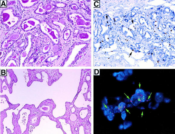
Postatrophic hyperplasia (PAH) of the prostate gland often demonstrates overlapping histological features with prostatic adenocarcinoma (PCA). These features include small acinar growth and enlarged nuclei with prominent nucleoli. Recent work has demonstrated that PAH is a proliferative, noninvoluting lesion. PAH is also histologically distinct from simple atrophy (SA), which has intermediate- to large-sized glands, minimal cytoplasm, and inconspicuous nuclei. However, despite overlapping features between PAH and PCA, high-grade prostatic intraepithelial neoplasm (HGPIN) is still considered the only direct neoplastic precursor to PCA. HGPIN resembles PCA in its topographic distribution, cytological appearance, and molecular alterations including chromosome 8p loss and chromosome 8 centromeric gain. To examine the hypothesis that PAH is the earliest histologically distinct precursor to HGPIN or PCA, the frequency, distribution, proliferative state, and chromosome 8 gain of benign prostate, SA, PAH, HGPIN, and PCA were analyzed. Forty radical prostatectomy specimens from men with clinically localized PCA were systematically analyzed. Proliferation was determined by Ki-67 immunohistochemistry (MIB-1) on formalin-fixed, paraffin-embedded tissue and quantified by digital image analysis from a total of 5,510 sample areas with benign, SA, PAH, HGPIN, and PCA. A tissue microarray was constructed to evaluate 8c gain using interphase fluorescence in situ hybridization. SA foci (n = 129) and PAH foci (n = 114) were identified in the 40 cases of which 74% (95 of 129) and 88% (100 of 114) were seen in the peripheral zone, respectively (P = 0.006). PAH and SA were identified adjacent to PCA in 28% (32 of 114) and 14% (18 of 129) of foci examined, respectively (P = 0.007). The median number of proliferating nuclei increased significantly from benign (1.20%), SA (2.67%), PAH (3.62%), HGPIN (6.14%), to PCA (12.00%) (P < 0.001). The median percentage of nuclei with more than three centromeric probe signals (chromosome 8c gain) for SA, HGPIN, PAH, and PCA were 2.1, 2.8, 4.0, and 6.0%, respectively, as compared to benign prostate with 1.3% (P = 0.006). In conclusion, the present study identified a strong topographic association between PAH and PCA. PAH is also seen often to be closely associated with chronic inflammation. Proliferation of PAH is significantly greater than benign prostatic epithelium and SA but less than HGPIN or PCA. Gain of 8c is significantly greater in PAH than benign prostate, SA, and even HGPIN. These findings demonstrate a strong association between PAH and PCA, supporting its role as a neoplastic precursor.
Figures
References
-
- Anton RC, Kattan MW, Chakraborty S, Wheeler TM: Postatrophic hyperplasia of the prostate: lack of association with prostate cancer. Am J Surg Pathol 1999, 23:932-936 - PubMed
-
- Franks L: Atrophy and hyperplasia in the prostate proper. J Pathol Bacteriol 1954, 68:617-621 - PubMed
-
- Liavag I: Atrophy and regeneration in the pathogenesis of prostatic carcinoma. Acta Pathol Microbiol Scand 1968, 73:338-350 - PubMed
-
- Qian J, Jenkins RB, Bostwick DG: Genetic and chromosomal alterations in prostatic intraepithelial neoplasia and carcinoma detected by fluorescence in situ hybridization. Eur Urol 1999, 35:479-483 - PubMed
-
- Qian J, Jenkins RB, Bostwick DG: Determination of gene and chromosome dosage in prostatic intraepithelial neoplasia and carcinoma. Anal Quant Cytol Histol 1998, 20:373-380 - PubMed
Publication types
MeSH terms
Substances
Grants and funding
LinkOut - more resources
Full Text Sources
Other Literature Sources
Medical
