. 2001 May;158(5):1775-84.
doi: 10.1016/S0002-9440(10)64133-8.

Regulation of gap junctional communication by a pro-inflammatory cytokine in cystic fibrosis transmembrane conductance regulator-expressing but not cystic fibrosis airway cells

Affiliations

Regulation of gap junctional communication by a pro-inflammatory cytokine in cystic fibrosis transmembrane conductance regulator-expressing but not cystic fibrosis airway cells

M Chanson et al. Am J Pathol. 2001 May.

Abstract

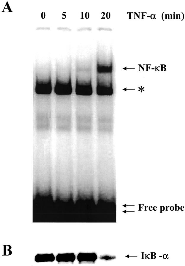
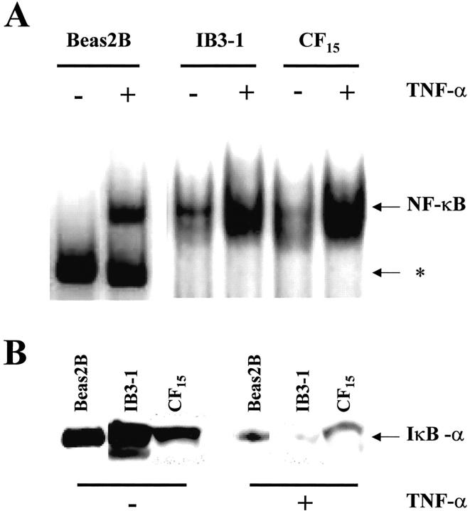
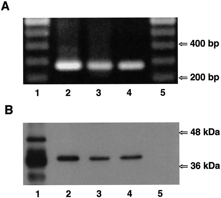
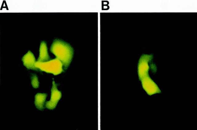
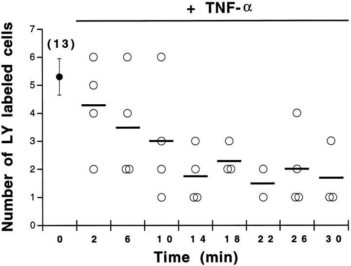
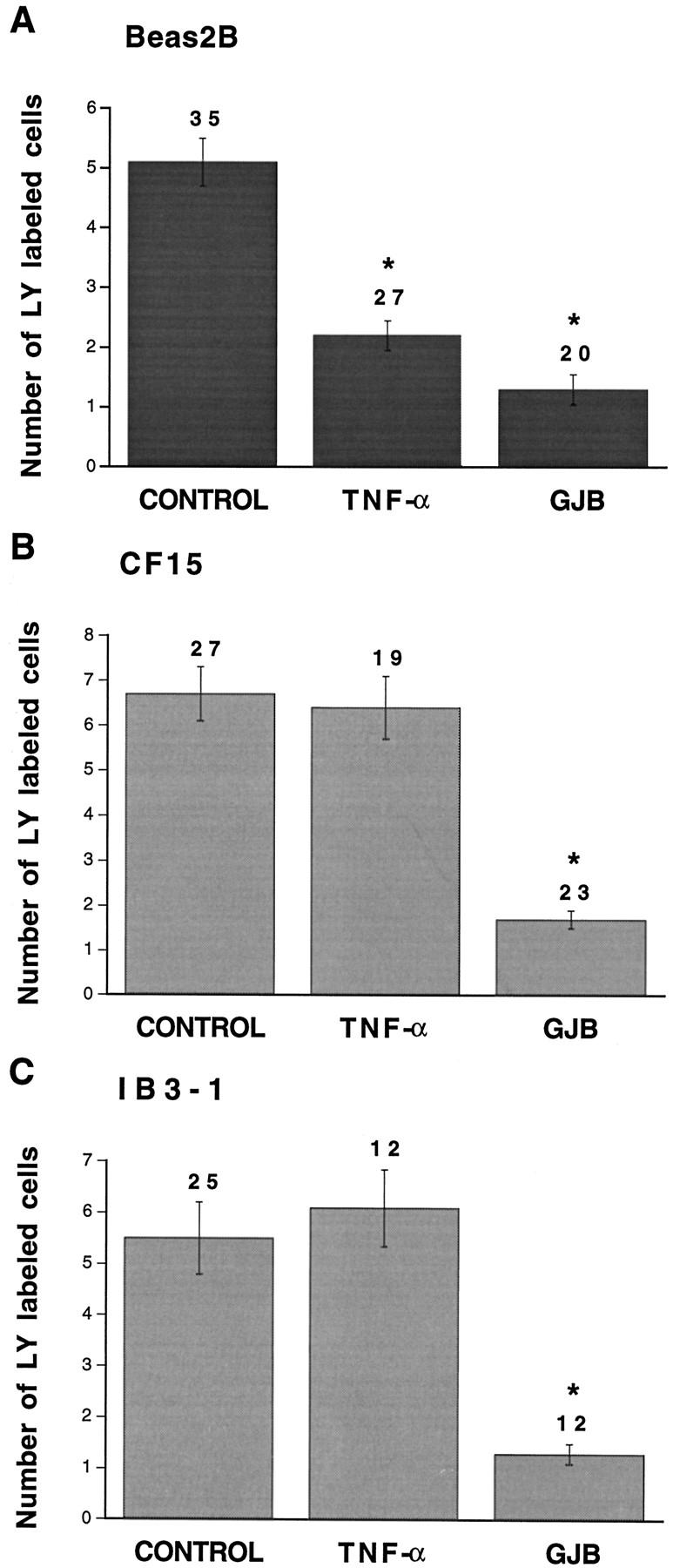
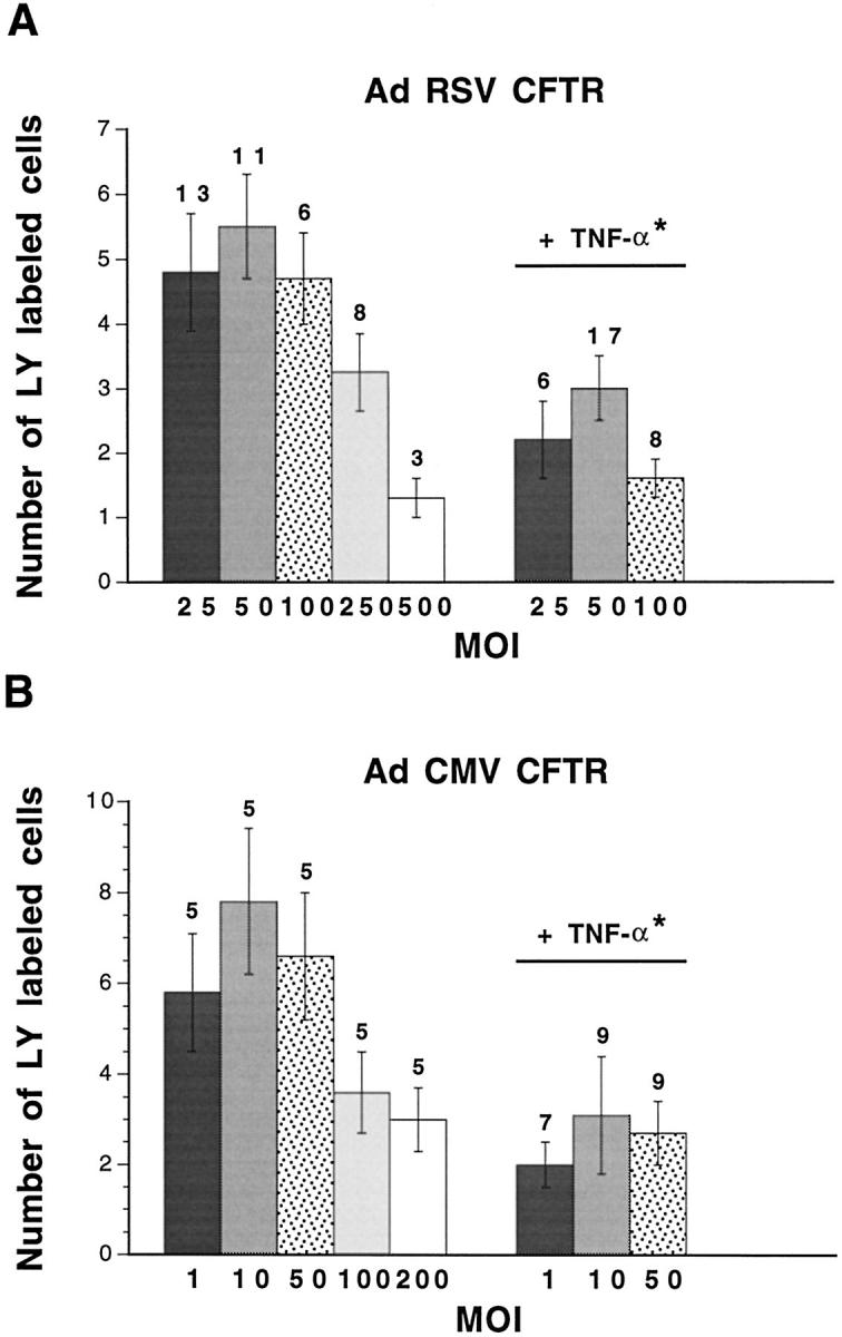
Airway inflammation is orchestrated by cell-cell interactions involving soluble mediators and cell adhesion molecules. Alterations in the coordination of the multicellular process of inflammation may play a major role in the chronic lung disease state of cystic fibrosis (CF). The aim of this study was to determine whether direct cell-cell interactions via gap junctional communication is affected during the inflammatory response of the airway epithelium. We have examined the strength of intercellular communication and the activation of nuclear factor-kappaB (NF-kappaB) in normal (non-CF) and CF human airway cell lines stimulated with tumor necrosis factor-alpha (TNF-alpha). TNF-alpha induced maximal translocation of NF-kappaB into the nucleus of non-CF as well as CF airway cells within 20 minutes. In non-CF cells, TNF-alpha progressively decreased the extent of intercellular communication. In contrast, gap junctional communication between CF cells exposed to TNF-alpha remained unaltered. CF results from mutations of the cystic fibrosis transmembrane conductance regulator (CFTR) gene. Interestingly, transfer of wild-type CFTR into CF cells by adenovirus-mediated infection was associated with the recovery of TNF-alpha-induced uncoupling. These results suggest that expression of functional CFTR is necessary for regulation of gap junctional communication by TNF-alpha. Gap junction channels close during the inflammatory response, therefore limiting the intercellular diffusion of signaling molecules, and thereby the recruitment of neighboring cells. Defects in this mechanism may contribute to the excessive inflammatory response of CF airway epithelium.


Figures

Figure 1.
Figure 1.
Time-course of NF-κB translocation and IκBα degradation in non-CF Beas2B cells exposed to TNF-α. A: EMSA analysis of NF-κB binding activity in nuclear extracts from Beas2B cells treated for the indicated time with 100 U/ml TNF-α. TNF-α induced the increase in NF-κB binding activity in nuclear extracts in a time-dependent manner. Maximal nuclear translocation was reached after 20 minutes in the presence of TNF-α. The single arrow corresponds to binding of NF-κB to the probe, and the double arrow indicates the free probe. Nonspecific binding of the NF-κB probe can also be seen on the EMSAs (asterisk). Competition with 100× excess of cold competitors (see Materials and Methods) abolished binding of NF-κB to the probe but not the nonspecific binding. B: Detection of IκBα by Western blot analysis of cytoplasmic extracts from Beas2B cells exposed to TNF-α. TNF-α rapidly induced the degradation of IκBα, in parallel to NF-κB translocation. The arrow indicates the IκBα band at ∼36 kd.
Figure 2.
Figure 2.
A: NF-κB translocation and IκBα degradation in non-CF and CF airway cells exposed to TNF-α. In all cell lines, EMSA analysis shows that TNF-α evoked maximal translocation of NF-κB within 20 minutes. Note nonspecific binding of the NF-κB probe in EMSAs on Beas2B cells (asterisk). B: The parallel degradation of IκBα elicited by TNF-α in Beas2B, IB3-1, and CF15 cells was detected by Western blot experiments after 20 minutes of cell exposure to TNF-α. The arrow indicates the IκBα band at ∼36 kd.
Figure 3.
Figure 3.
Expression of Cx43 in non-CF and CF airway cells. A: Reverse transcriptase-polymerase chain reaction was performed on mRNA isolated from Beas2B (lane 2), CF15 (lane 3), and IB3-1 (lane 4) cells using primer pairs specific for human Cx43. Amplification products of the expected sizes for Cx43 (285 bp) were detected in all non-CF and CF cell lines. Molecular markers are shown in lanes 1 and 5. B: Western blot analysis of Cx43 expression in non-CF and CF airway cells. The anti-Cx43 antibody revealed one band at ∼41 kd in cytosolic protein extracts from Beas2B (lane 2), CF15 (lane 3), and IB3-1 (lane 4) cells. Lane 1 corresponds to rat atrium samples that were used as positive controls. Several bands at 41 to 46 kd could be detected, corresponding to various phosphorylated forms of Cx43. The lower band likely corresponds to a degradation product of Cx43. Lane 5 corresponds to SKHep1 cells, which do not express Cx43, that were used as negative controls.
Figure 4.
Figure 4.
Effects of TNF-α on the extent of dye coupling between non-CF Beas2B cells. Under control conditions, lucifer yellow (LY) diffused from the microinjected cell to five to six neighbors (A). In contrast, the extent of LY diffusion was markedly decreased in the presence of 100 U/ml TNF-α (B). Scale bar, 15 μm.
Figure 5.
Figure 5.
Time course of TNF-α-induced dye uncoupling between non-CF cells. In the absence of TNF-α (time 0), the number of cells labeled with lucifer yellow (LY) averaged 5.3 ± 0.65 cells (n = 13 microinjections). Exposure of Beas2B cells to TNF-α was associated with a time-dependent decrease in the number of LY-labeled cells. Maximal uncoupling was reached within 20 minutes. Bars indicate the mean number of LY-labeled cells.
Figure 6.
Figure 6.
Quantitative evaluation of dye coupling in non-CF and CF airway cells exposed to TNF-α. Under control conditions, transfer of lucifer yellow (LY) was detected in Beas2B (A), CF15 (B), and IB3-1 (C) cells. Whereas TNF-α significantly decreased dye transfer in non-CF Beas2B cells, the pro-inflammatory mediator had no effect on intercellular communication between CF (CF15 and IB3-1) cells. In all cells lines, gap junction channel blockers (GJB) inhibited dye coupling. Asterisks indicate differences at P < 0.002 levels.
Figure 7.
Figure 7.
Dye coupling in CF15 cells infected with AdRSV CFTR or AdCMV CFTR. High MOIs of AdRSV CFTR (A) and of AdCMV CFTR (B) decreased lucifer yellow (LY) diffusion between CF15 cells. Lower MOIs of AdRSV CFTR (25 to 100) and of AdCMV CFTR (1 to 50) did not affect the normal extent of intercellular communication between CF15 cells. For the latter range of concentrations, exposure of the cells to TNF-α was associated with a significant reduction of gap junctional communication. Thus, difference at P < 0.001 (asterisk) was calculated for the pooled values obtained from 25 to 100 MOIs of AdRSV CFTR- or 1 to 50 MOIs of AdCMV CFTR-infected cells between control and TNF-α conditions.

Comment in

Similar articles

Cited by

References

    1. Welsh MJ, Tsui L-C, Boat TM, Beaudet AL: Cystic fibrosis. ed 7 Scriver CR Beaudet AL Sly WS Valle D eds. The Metabolic and Molecular Basis of Inherited Disease, 1995, :pp 3799-3876 McGraw-Hill, New York
    1. Schwiebert EM, Benos DJ, Egan ME, Stutts MJ, Guggino WB: CFTR is a conductance regulator as well as a chloride channel. Physiol Rev 1999, 79:S145-S166 - PubMed
    1. Tosi MF, Stark JM, Smith CW, Hamedani A, Gruenert DC, Infeld MD: Induction of ICAM-1 expression on human airway epithelial cells by inflammatory cytokines: effects on neutrophil-epithelial cell adhesion. Am J Respir Cell Mol Biol 1992, 7:214-221 - PubMed
    1. Bonfield TM, Panuska JR, Konstan MW, Hilliard KA, Hilliard JB, Ghnaim H, Berger M: Inflammatory cytokines in cystic fibrosis lungs. Am J Respir Crit Care Med 1995, 152:2111-2118 - PubMed
    1. Noah TL, Black HR, Cheng PW, Wood RE, Leigh MW: Nasal and bronchoalveolar lavage fluid cytokines in early cystic fibrosis. J Infect Dis 1997, 175:638-647 - PubMed

Publication types

MeSH terms