. 2001 May;158(5):1833-42.
doi: 10.1016/S0002-9440(10)64139-9.

Increased osteoblast apoptosis in apert craniosynostosis: role of protein kinase C and interleukin-1

Affiliations

Increased osteoblast apoptosis in apert craniosynostosis: role of protein kinase C and interleukin-1

J Lemonnier et al. Am J Pathol. 2001 May.

Abstract

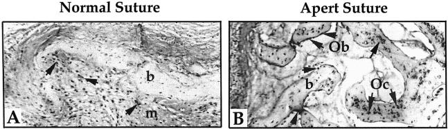
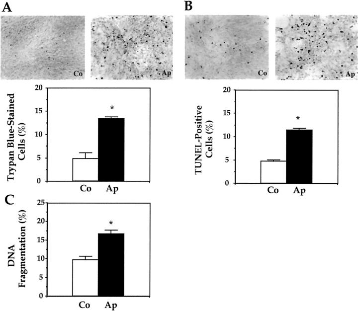
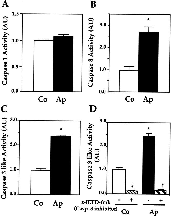
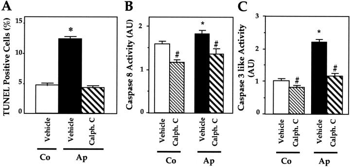
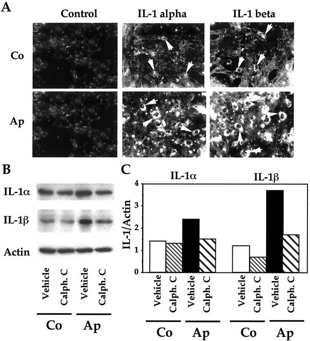
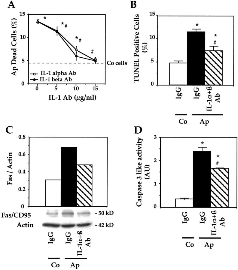
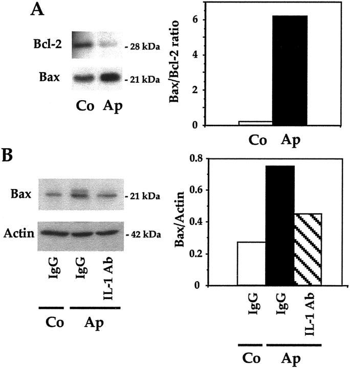
Apert syndrome is an autosomal dominant disorder characterized by premature cranial ossification resulting from fibroblast growth factor receptor-2 (FGFR-2)-activating mutations. We have studied the effects of the prominent S252W FGFR-2 Apert mutation on apoptosis and the underlying mechanisms in human mutant osteoblasts. In vivo analysis of terminal deoxynucleotidyl transferase-mediated nick-end labeling revealed premature apoptosis of mature osteoblasts and osteocytes in the Apert suture compared to normal coronal suture. In vitro, mutant osteoblasts showed increased apoptosis, as demonstrated by terminal deoxynucleotidyl transferase-mediated nick-end labeling analysis, trypan blue staining, and DNA fragmentation. Mutant osteoblasts also showed increased activity of caspase-8 and effector caspases (-3, -6, -7) constitutively. This was related to protein kinase C activation because the selective protein kinase C inhibitor calphostin C inhibited caspase-8, effector caspases, and apoptosis in mutant osteoblasts. Apert osteoblasts also showed increased expression of interleukin (IL)-1alpha, IL-1beta, Fas, and Bax, and decreased Bcl-2 levels. Specific neutralizing anti-IL-1 antibody reduced Fas levels, Bax expression, effector caspases activity, and apoptosis in mutant cells. Thus, the Apert S252W FGFR-2 mutation promotes apoptosis in human osteoblasts through activation of protein kinase C, overexpression of IL-1 and Fas, activation of caspase-8, and increased Bax/Bcl-2 levels, leading to increased effector caspases and DNA fragmentation. This identifies a complex FGFR-2 signaling pathway involved in the premature apoptosis induced by the Apert S252W FGFR-2 mutation in human calvaria osteoblasts.


Figures

Figure 1.
Figure 1.
The Apert S252W FGFR-2 mutation induces premature osteoblast apoptosis in the human suture. Normal (A) and Apert (B) coronal sutures were prepared for TUNEL analysis. The Apert suture shows numerous TUNEL-positive mature osteoblasts (Ob) along the bone trabeculae and TUNEL-positive osteocytes (Oc) in the bone (b) matrix (arrows) whereas only mesenchymal (m) cells were found to be TUNEL-positive in the normal suture. Original magnification, ×125.
Figure 2.
Figure 2.
Increased in vitro apoptosis induced by the S252W FGFR-2 mutation in Apert mutant osteoblasts. Apert (Ap) and control (Co) cells were stained with trypan blue (A) or TUNEL (B) and the number of trypan blue-stained or TUNEL-positive cells was counted. C: DNA fragmentation was determined as indicated in Materials and Methods. The data are the mean ± SEM of four values. *, Significant difference with Co cells (P < 0.05).
Figure 3.
Figure 3.
Increased caspase-8- and -3-like activity in mutant osteoblasts. Caspase-1 (A), caspase-8 (B), and caspase-3, -6, and -7 (caspase-like) (C) were determined in Co and Ap cells. Caspase-3, -6, and -7 activity was also determined in the absence or presence of caspase-8 inhibitor (z-IETD-fmk) (D). Ap cells showed increased caspase-8 and caspase-3 activities, and the latter was inhibited by the specific caspase-8 inhibitor. The data are the mean ± SEM of four values. *, P < 0.05 versus Co cells; #, P < 0.05 versus Co or Ap cells untreated with the caspase-8 inhibitor.
Figure 4.
Figure 4.
Role of PKC in caspase-8- and -3-like activity and apoptosis in mutant osteoblasts. Mutant Apert (Ap) cells were treated with 2 μmol/L calphostin C, a specific PKC inhibitor, and TUNEL-positive apoptotic cells (A), caspase-8 (B), and caspase-3, -6, and -7 (caspase-like) activity (C) were determined. The data are the mean ± SEM of four values. *, P < 0.05 versus Co cells; #, P < 0.05 versus vehicle-treated Co or Ap cells.
Figure 5.
Figure 5.
Role of PKC in the increased IL-1α and IL-1β expression in mutant osteoblasts. A: Apert (Ap) and control (Co) cells were immunostained for IL-1α and IL-1β using specific antibodies or IgG (control). Strong IL-1α and IL-1β immunostaining was found in Ap cells compared to Co cells (arrows). Western blot analysis (B) and densitometric analysis (C) show that the IL-1α and IL-1β overexpression in Ap cells was suppressed by the PKC inhibitor calphostin C (2 μmol/L).
Figure 6.
Figure 6.
Role of IL-1 and Fas in the increased apoptosis in mutant osteoblasts. A: Apert (Ap) cells were treated with anti-IL-Iα or anti-IL-1β antibodies at different doses and the number of trypan blue-stained cells was recorded. The dotted bar indicates the normal level in control (Co) cells. B: Ap cells were treated with neutralizing IL-1α and IL-1β antibodies (15 μg/ml each) or IgG, and the number of TUNEL-positive cells was counted. In parallel experiments, Fas protein levels were determined by Western blot analysis and the data recorded by densitometric analysis were corrected for β-actin (C). Caspase-3, -6, and -7 (caspase-like) activity was determined in the same culture conditions (D). The data are the mean ± SEM of four values. *, P < 0.05 versus Co IgG-treated cells; #, P < 0.05 versus Ap IgG-treated cells.
Figure 7.
Figure 7.
Role of Bax and Bcl-2 in apoptosis in mutant osteoblasts. The levels of the pro-apoptotic protein Bax and the anti-apoptotic protein Bcl-2 were determined by Western blot analysis in Apert (Ap) and control (Co) cells in basal culture conditions, the bands were scanned and the ratio of Bax/Bcl-2 was determined (A). Bax protein levels were determined in Ap cells in the presence of specific neutralizing IL-1α and IL-1β antibodies (15 μg/ml each) or IgG, and the levels were corrected for β-actin (B).

References

    1. Muenke M, Schell U: Fibroblast-growth-factor receptor mutations in human skeletal disorders. Trends Genet 1995, 11:308-313 - PubMed
    1. Naski MC, Ornitz DM: FGF signaling in skeletal development. Front Biosci 1998, 3:D781-D794 - PubMed
    1. Burke D, Wilkes D, Blundell TL, Malcolm S: Fibroblast growth factor receptors: lessons from the genes. Trends Biochem Sci 1998, 23:59-62 - PubMed
    1. Hehr U, Muenke M: Craniosynostosis syndromes: from genes to premature fusion of skull bones. Mol Genet Metab 1999, 68:139-151 - PubMed
    1. Mangasarian K, Li Y, Mansukhani A, Basilico C: Mutation associated with Crouzon syndrome causes ligand-independent dimerization and activation of FGF receptor-2. J Cell Physiol 1997, 172:117-125 - PubMed

MeSH terms