. 2001 Jul 6;276(27):24965-70.
doi: 10.1074/jbc.M101161200. Epub 2001 May 3.

Inositol hexakisphosphate kinase 2 mediates growth suppressive and apoptotic effects of interferon-beta in ovarian carcinoma cells

Affiliations

Inositol hexakisphosphate kinase 2 mediates growth suppressive and apoptotic effects of interferon-beta in ovarian carcinoma cells

B H Morrison et al. J Biol Chem. .

Abstract

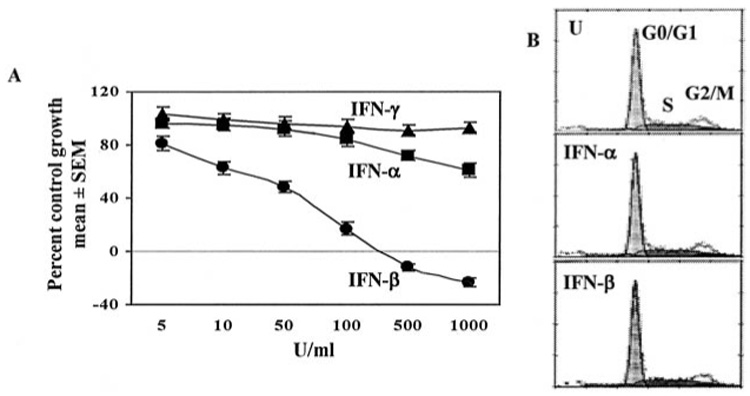
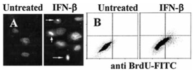
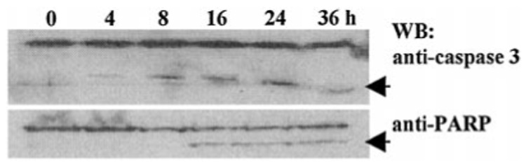
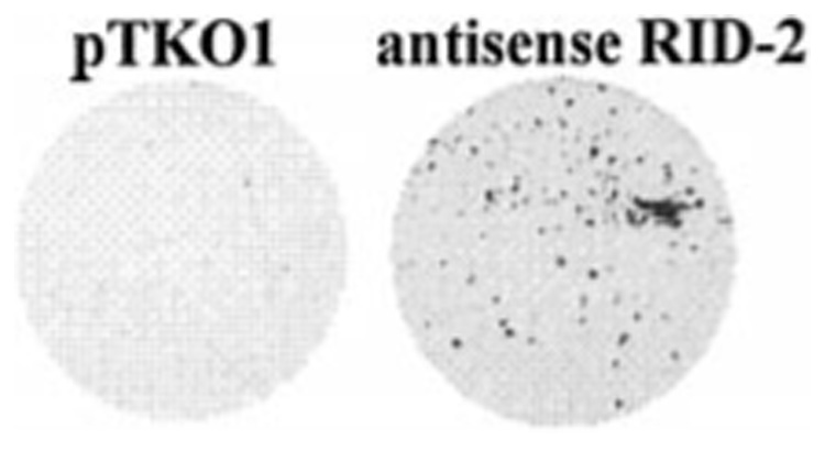
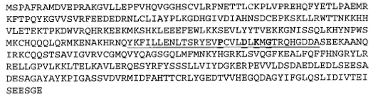
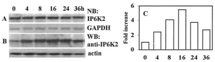
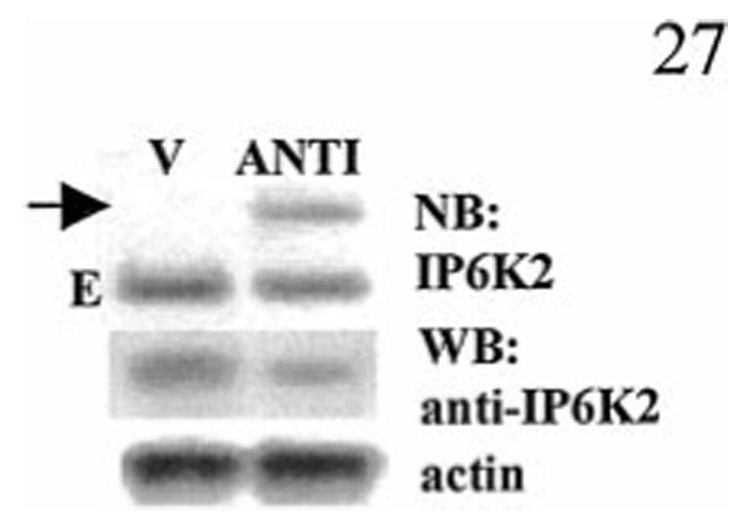
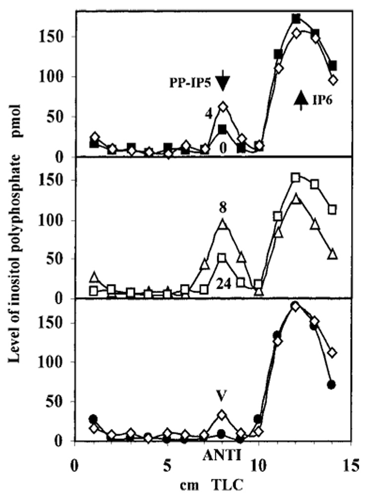
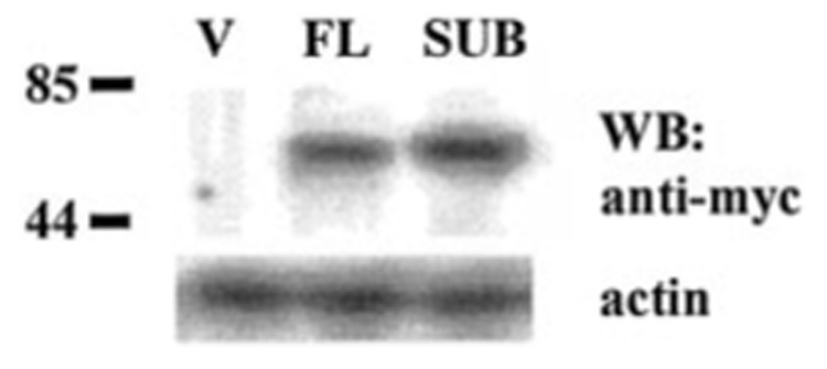
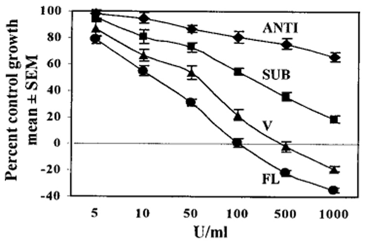
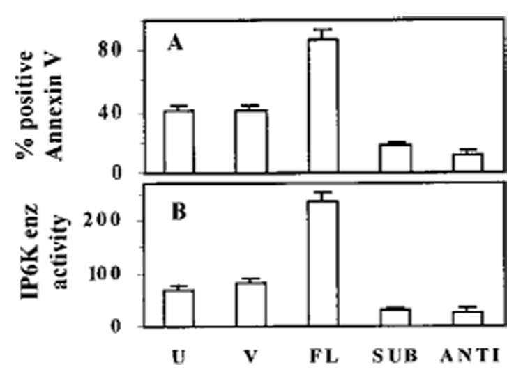
Interferons (IFNs) regulate the expression of genes that mediate their antiviral, antitumor, and immunomodulatory actions. We have previously shown that IFN-beta suppresses growth of human ovarian carcinoma xenografts in vivo and induces apoptosis of ovarian carcinoma cells in vitro. To investigate mechanisms of IFN-beta-induced apoptosis we employed an antisense technical knockout approach to identify gene products that mediate cell death and have isolated several regulators of interferon-induced death (RIDs). In this investigation, we have characterized one of the RIDs, RID-2. Sequence analysis revealed that RID-2 was identical to human inositol hexakisphosphate kinase 2 (IP6K2). IP6K2 is post-transcriptionally induced by IFN-beta in ovarian carcinoma cells. A mutant IP6K2 with substitutions in the putative inositol phosphate binding domain abrogates IFN-beta-induced apoptosis. These studies identify a novel function for IP6K2 in cell growth regulation and apoptosis.


Figures

FIG. 1.
FIG. 1.. IFN-β but not IFN-α or IFN-γ induces ovarian carcinoma cell death
A, NIH-OVCAR-3 cells were grown in the presence of 5–1000 units/ml IFN-β, IFN-α, or IFN-γ. After 7 days cells were fixed and stained with sulforhodamine B. Absorbance (570 nm) of bound dye was measured and expressed as the percent of untreated controls. Each data point represents mean ± S.E. of eight replicates. Values on the negative scale indicate death of initially plated cells. B, cells were left untreated (U) or treated with IFN-β or IFN-α (200 units/ml) for 2 days. Cells were stained with propidium iodide and subjected to flow cytometry and cell cycle analysis (MultiPass).
FIG. 2.
FIG. 2.. IFN-β induces apoptosis in NIH-OVCAR-3 cells
A, cells were treated with IFN-β (200 units/ml) for 2 days. Condensed, DAPI-stained apoptotic nuclei are visible (arrows). B, TUNEL assay. Cells were treated with IFN-β as above, labeled with bromo-dUTP using terminal deoxynucleotidyltransferase, and stained with FITC-conjugated anti-bromodeoxyuridine monoclonal antibody (x axis) and PI (y axis). The percentage of FITC-positive cells (in upper right and lower right quadrants) was determined by flow cytometry.
FIG. 3.
FIG. 3.. IFN-β activates caspase 3 during apoptosis
Western blots (WB) of caspase 3 (upper) and PARP (lower). Lysates (40 µg of protein) of NIH-OVCAR-3 cells that were treated with 0–36 h of IFN-β (200 units/ml) are shown. Caspase 3 cleavage products (top arrow) appear earlier than PARP cleavage products (bottom arrow).
FIG. 4.
FIG. 4.. Protection of NIH-OVCAR-3 cells by antisense IP6K2 episome
Cells were electroporated with 20 µg of pTKO1 (left) or antisense IP6K2 episomes (right) and selected for 4 weeks with IFN-β and hygromycin B. Surviving cells were fixed and stained with sulfor-hodamine B.
FIG. 5.
FIG. 5.. Identification of RID-2 as human IP6K2
The amino acid sequence of IP6K2, GenBank™ accession number AAF15057, is shown. Putative IPBD is underlined with most highly conserved residues shown in bold.
FIG. 6.
FIG. 6.. Effects of IFN-β on IP6K2 expression
(A), Northern blot (NB) analysis. Total RNA (40 µg) derived from NIH-OVCAR-3 cells after treatment with IFN-β (200 units/ml for 0–36 h) are shown; blots were hybridized with IP6K2 cDNA probe and glyceraldehyde-3-phosphate dehydrogenase as control. B, Western blot (WB) analysis of whole cell lysates (70 µg) prepared after IFN-β treatment as above. Blots were probed with anti-IP6K2 antibody and anti-actin as control. C, densitometric quantitation of IP6K2 protein levels from B. The x axis indicates time of IFN-β exposure (0–36 h), and the y axis indicates band intensity normalized to actin.
FIG. 7.
FIG. 7.. Expression of antisense IP6K2 mRNA reduces IP6K2 protein levels
Upper panel, Northern blot (NB) analysis of pools (~100 clones each) of NIH-OVCAR-3 cells stably transfected with vector (V) or antisense IP6K2 (ANTI). The arrow indicates the position of mRNA in transgene-expressing cells. E indicates endogenous mRNA. Lower panel, IP6K2 Western blot (WB) of same cells and actin as control.
FIG. 8.
FIG. 8.. Time course of IP6K activity in IFN-β-stimulated NIH-OVCAR-3 cells
Cells were treated with IFN-β (200 units/ml). Numbers next to curves in top and middle panels (0, 4, 8, and 24) indicate treatment duration (h). IP6K enzymatic activity in whole cell homogenates was determined using [³H]IP6 as substrate in kinase reactions in vitro and detected by PEI-TLC. Total radioactivity contained in the [³H]PP-IP5 product, represented as AUC, for IFN-β-treated cells at various time points was 70 (0 h), 120 (4 h), 209 (8 h), and 101 (24 h). Lower panel, empty vector (V) and antisense IP6K2 (ANTI)-expressing cells. AUC for V cells was 72, and for ANTI cells it was 32. Arrows indicate Rf for PP-IP5 and IP6.
FIG. 9.
FIG. 9.. Expression of mutant IP6K2 in NIH-OVCAR-3 cells
Lysates (80 µg) from cell lines were separated on SDS-polyacrylamide gel electrophoresis and probed with antibody directed against Myc tag (Invitrogen). Labels indicate plasmids transfected into cells. V, pCXN2myc vector; FL, full-length IP6K2. Predicted molecular mass (kDa) of tagged IP6K2 proteins is 51.7 for FL and 51.5 for SUB. Western blots (WB) were stripped and reprobed with actin as control. Tagged proteins were detected in FL and SUB but not in vector (V) cells.
FIG. 10.
FIG. 10.. Effect of IFN-β on growth of NIH-OVCAR-3 cells expressing SUB mutant
Growth assays were performed as described in legend for Fig. 1. Each point represents mean ± S.E. of eight replicates. Notations are similar to those in Fig. 9. Cells expressing ANTI IP6K2 mRNA were most resistant to IFN-β; cells overexpressing full-length IP6K2 were most sensitive.
FIG. 11.
FIG. 11.. Apoptosis and IP6K enzymatic activity in transfected cell lines
A, U indicates untransfected NIH-OVCAR-3 cells. Other notations are similar to those in Fig. 9. Cell lines were treated with IFN-β (200 units/ml) and subjected to annexin staining. The percentage of apoptotic cells was determined by flow cytometry. B, in parallel, IP6K enzymatic activity was determined in whole cell lysates. IP6K enzyme activity is represented as total area under PP-IP-5 curve. Annexin V and enzymatic data are shown as mean ± S.E. of three separate experiments.

References

    1. Gresser I, Bourali C, Levy JP, Fontaine-Brouty-Boye D, Thomas MT. Proc. Natl. Acad. Sci. U. S. A. 1969;63:51–57. - PMC - PubMed
    1. Evinger M, Rubinstein M, Pestka S. Arch. Biochem. Biophys. 1981;210:319–329. - PubMed
    1. Kalvakolanu DV. Histol. Histopathol. 2000;15:523–537. - PubMed
    1. Stark GR, Kerr IM, Williams BR, Silverman RH, Schreiber RD. Ann. Rev. Biochem. 1998;67:227–264. - PubMed
    1. Darnell JEJ. Science. 1997;277:1630–1635. - PubMed

Publication types

MeSH terms