. 2001 Jun;21(11):3750-62.
doi: 10.1128/MCB.21.11.3750-3762.2001.

Activation of the Ral and phosphatidylinositol 3' kinase signaling pathways by the ras-related protein TC21

Affiliations

Activation of the Ral and phosphatidylinositol 3' kinase signaling pathways by the ras-related protein TC21

M Rosário et al. Mol Cell Biol. 2001 Jun.

Abstract

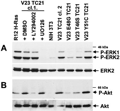
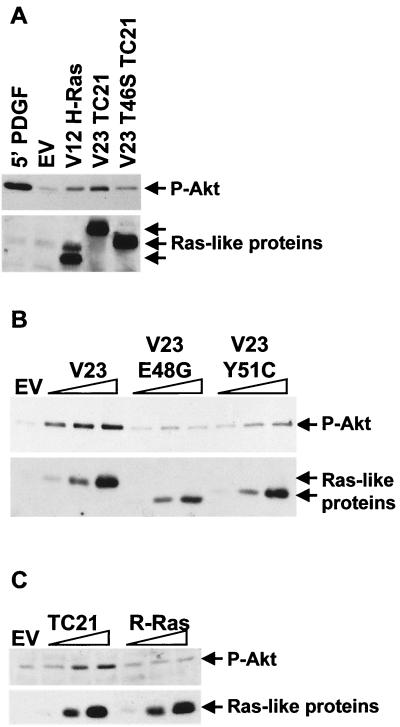
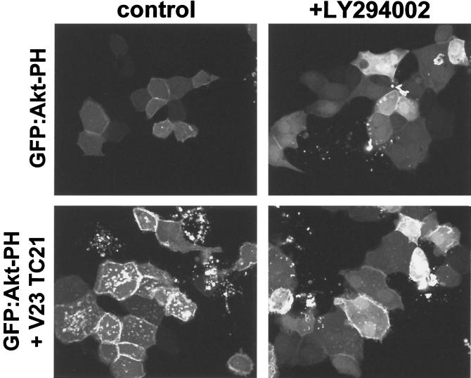
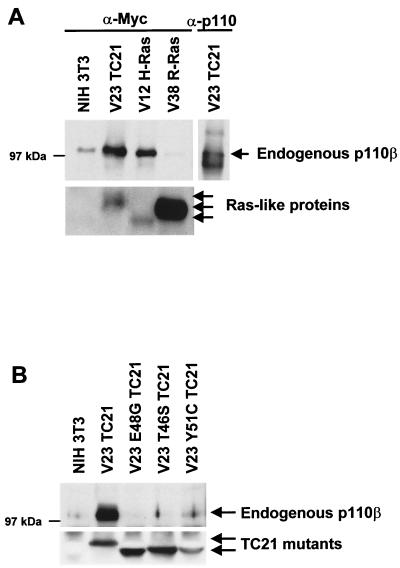
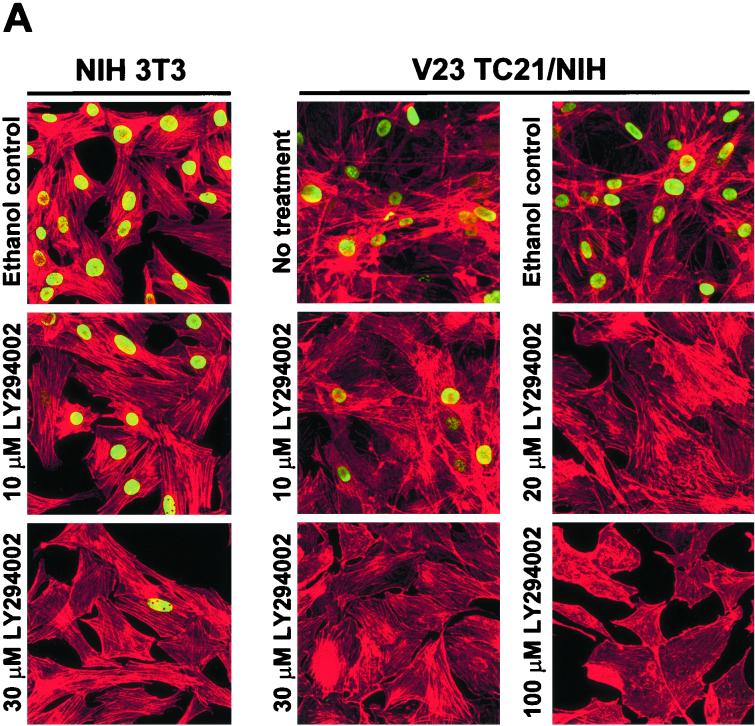
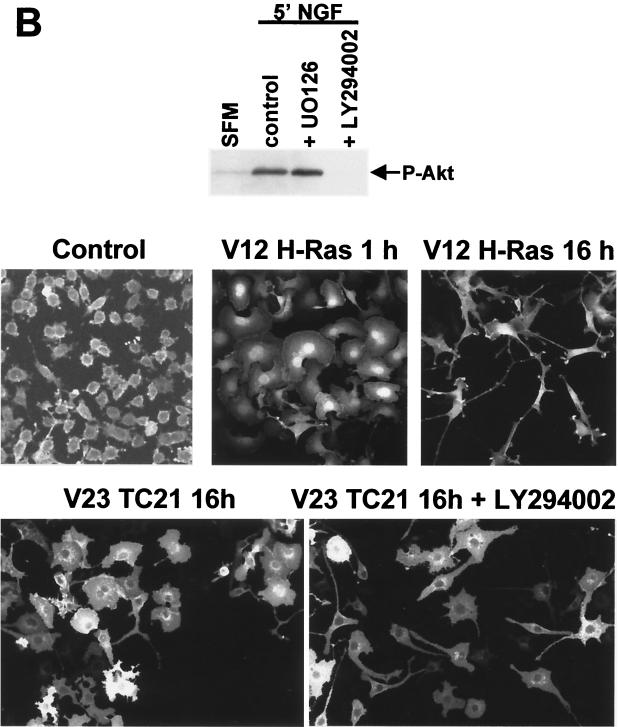
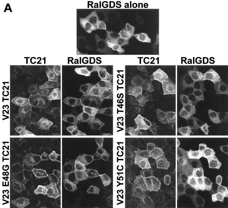
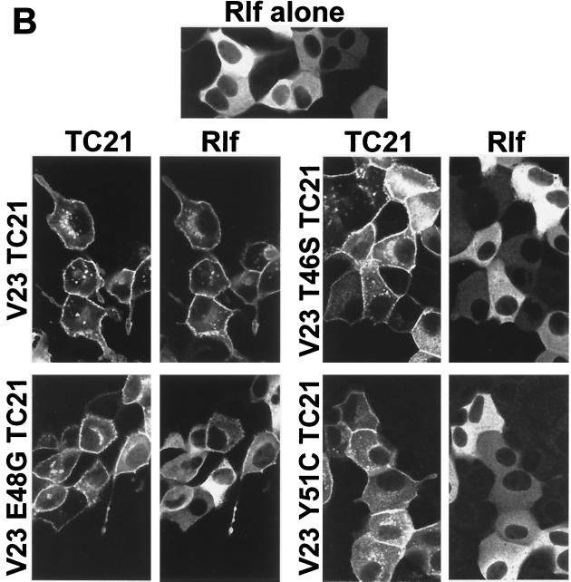
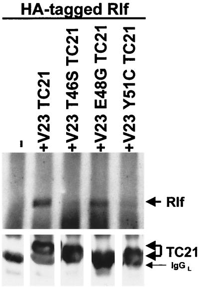
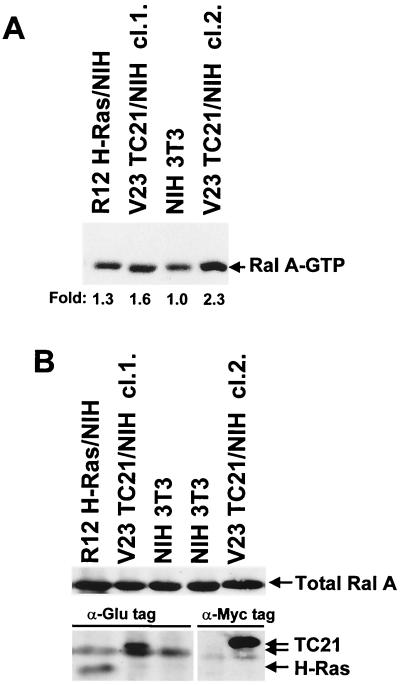
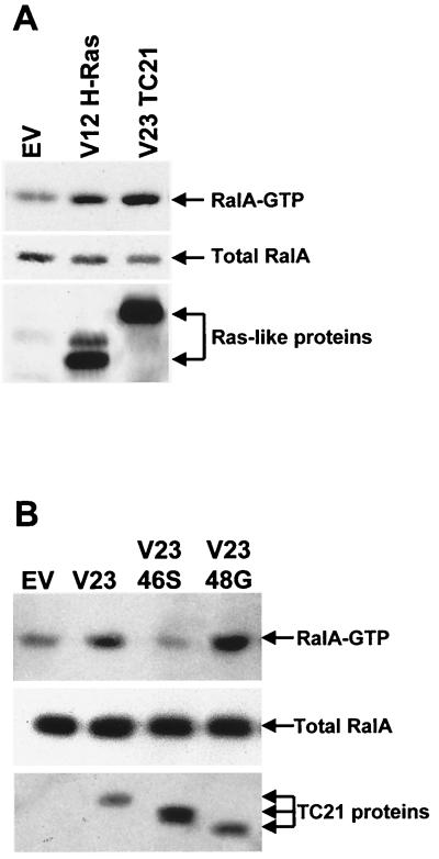
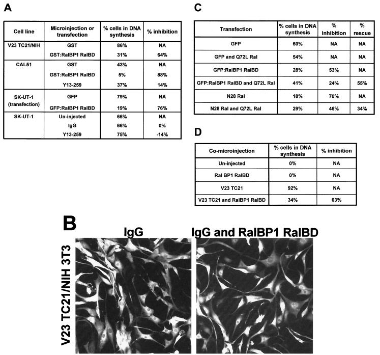
TC21 is a member of the Ras superfamily of small GTP-binding proteins that, like Ras, has been implicated in the regulation of growth-stimulating pathways. We have previously identified the Raf/mitogen-activated protein kinase pathway as a direct TC21 effector pathway required for TC21-induced transformation (M. Rosário, H. F. Paterson, and C. J. Marshall, EMBO J. 18:1270-1279, 1999). In this study we have identified two further effector pathways for TC21, which contribute to TC21-stimulated transformation: the phosphatidylinositol 3' kinase (PI-3K) and Ral signaling pathways. Expression of constitutively active TC21 leads to the activation of Ral A and the PI-3K-dependent activation of Akt/protein kinase B. Strong activation of the PI-3K/Akt pathway is seen even with very low levels of TC21 expression, suggesting that TC21 may be a key small GTPase-regulator of PI-3K. TC21-induced alterations in cellular morphology in NIH 3T3 and PC12 cells are also PI-3K dependent. On the other hand, activation of the Ral pathway by TC21 is required for TC21-stimulated DNA synthesis but not transformed morphology. We show that inhibition of Ral signaling blocks DNA synthesis in human tumor cell lines containing activating mutations in TC21, demonstrating for the first time that this pathway is required for the proliferation of human tumor cells. Finally, we provide mechanisms for the activation of these pathways, namely, the direct in vivo interaction of TC21 with guanine nucleotide exchange factors for Ral, resulting in their translocation to the plasma membrane, and the direct interaction of TC21 with PI-3K. In both cases, the effector domain region of TC21 is required since point mutations in this region can interfere with activation of downstream signaling.


Figures

FIG. 1
FIG. 1
Activation of the ERK/MAP kinase and Akt/PI-3K pathways in TC21-transformed cells. (A) NIH 3T3 cell lines stably expressing oncogenic TC21, H-Ras, or TC21 proteins containing mutations in the effector domain and untransformed NIH 3T3 were transferred to serum-free medium for 4 h. TC21-transformed cells were also exposed either to 25 μM LY294002, 20 μM UO126 (a MEK inhibitor), or the equivalent volume of ethanol carrier for 2 h prior harvesting. Whole-cell lysates were separated by sodium dodecyl sulfate-polyacrylamide gel electrophoresis (SDS-PAGE), and specific antibodies were used to detect phosphorylated ERK1 and ERK2 as well as total ERK2. Two independently isolated clones of TC21-transformed cells (cl.1 and cl.2) are shown. (B) The transformed NIH 3T3 cell lines were also analyzed for levels of phosphorylated Akt/PKB and total Akt/PKB in the presence of 50 μM LY294002, 20 μM UO126, or the equivalent volume of dimethyl sulfoxide (DMSO) carrier added for 2 h prior to harvesting as described above. Antibodies against the phosphorylated Ser 473 or Thr 308 sites in Akt/PKB gave similar results.
FIG. 2
FIG. 2
Activation of endogenous Akt/PKB by TC21. NIH 3T3 cells were transiently transfected with fixed or increasing amounts of expression vectors for Myc-tagged constitutively active forms of H-Ras, TC21, the TC21 effector mutants, R-Ras, or an empty-vector control (EV). The cells were harvested 20 h posttransfection following a 4-h serum starvation. One of the empty-vector transfections was stimulated with 25 ng of platelet-derived growth factor (PDGF) per ml for 5 min prior to harvesting. Whole-cell lysates equilibrated for protein levels were separated by SDS-PAGE. Specific anti-phosphorylated Akt/PKB and anti-Myc antibodies were used to detect the active Akt/PKB and the Ras-like proteins, respectively.
FIG. 3
FIG. 3
Translocation of the PH domain of Akt/PKB to the plasma membrane by TC21. Confluent MDCK cells were microinjected with 10 μg of an expression vector for the GFP:Akt PH domain fusion protein per ml alone or in conjunction with 50 μg of an expression vector for the Myc-tagged V23 TC21 per ml. LY294002 (25 μM) or the equivalent volume of ethanol was added to the medium just prior to microinjection. Cells were fixed 8 h after microinjection. GFP fluorescence is shown.
FIG. 4
FIG. 4
Interaction of TC21 with PI-3K. Myc-tagged oncogenic TC21, R-Ras, and H-Ras (A) or Myc-tagged constitutively active TC21 proteins containing specific mutations in the effector domain (B) were immunoprecipitated from NIH 3T3 cell lines stably expressing these proteins and grown in medium supplemented with 5% serum. Coimmunoprecipitating endogenous p110 was detected after SDS-PAGE by immunoblotting with specific antibodies.
FIG. 5
FIG. 5
TC21-induced transformation of NIH 3T3 cells (A) and morphological alteration of PC12 cells (B) are dependent on PI-3K activation. (A) V23 TC21-transformed NIH 3T3 or parental untransformed NIH 3T3 cells were seeded on several plastic dishes in DMEM supplemented with 5% serum. The cells were exposed to various concentrations of LY294002 or to the equivalent volume of the ethanol carrier for 42 h and allowed to incorporate BrdU during the last 15 h. Samples were then fixed and stained for the incorporated BrdU (shown in green) and for polymerized actin (shown in red). (B) PC12 cells were seeded on laminin-coated plates in medium containing 5% fetal calf serum and 10% horse serum. The medium was diluted 1:4 with serum-free medium 24 h after seeding. At 48 h after seeding, the cells were microinjected with 25 μg of an expression vector for Myc-tagged oncogenic TC21 or H-Ras per ml. LY294002 (25 μM) was added to a second duplicate plate of cells after injection. The cells were fixed 15 h after injection and stained for TC21 or H-Ras expression using anti-Myc antibodies. Levels of phosphorylated Akt/PKB in duplicate plates exposed to 25 μM LY294002, 20 μM UO126, or the equivalent volume of dimethyl sulfoxide carrier for 4 h prior to stimulation with 100 ng of NGF per ml for 5 min are shown.
FIG. 5
FIG. 5
TC21-induced transformation of NIH 3T3 cells (A) and morphological alteration of PC12 cells (B) are dependent on PI-3K activation. (A) V23 TC21-transformed NIH 3T3 or parental untransformed NIH 3T3 cells were seeded on several plastic dishes in DMEM supplemented with 5% serum. The cells were exposed to various concentrations of LY294002 or to the equivalent volume of the ethanol carrier for 42 h and allowed to incorporate BrdU during the last 15 h. Samples were then fixed and stained for the incorporated BrdU (shown in green) and for polymerized actin (shown in red). (B) PC12 cells were seeded on laminin-coated plates in medium containing 5% fetal calf serum and 10% horse serum. The medium was diluted 1:4 with serum-free medium 24 h after seeding. At 48 h after seeding, the cells were microinjected with 25 μg of an expression vector for Myc-tagged oncogenic TC21 or H-Ras per ml. LY294002 (25 μM) was added to a second duplicate plate of cells after injection. The cells were fixed 15 h after injection and stained for TC21 or H-Ras expression using anti-Myc antibodies. Levels of phosphorylated Akt/PKB in duplicate plates exposed to 25 μM LY294002, 20 μM UO126, or the equivalent volume of dimethyl sulfoxide carrier for 4 h prior to stimulation with 100 ng of NGF per ml for 5 min are shown.
FIG. 6
FIG. 6
Plasma membrane translocation of RalGDS and Rlf by V23 and V23 E48G TC21. Confluent MDCK cells were microinjected with 20 μg of an expression vector for a GFP:wild-type RalGDS fusion protein per ml (A) or 50 μg of an expression vector for HA-tagged wild-type Rlf per ml (B) alone or in conjunction with expression vectors for Myc-tagged V23 TC21 wild-type or effector mutant proteins (25 and 50 μg/ml, respectively). Cells were fixed and stained 14 h after microinjection for Myc-tagged TC21 or HA-tagged Rlf using specific antibodies. GFP fluorescence is shown for the GFP:RalGDS fusion protein. Samples were analyzed by confocal microscopy.
FIG. 6
FIG. 6
Plasma membrane translocation of RalGDS and Rlf by V23 and V23 E48G TC21. Confluent MDCK cells were microinjected with 20 μg of an expression vector for a GFP:wild-type RalGDS fusion protein per ml (A) or 50 μg of an expression vector for HA-tagged wild-type Rlf per ml (B) alone or in conjunction with expression vectors for Myc-tagged V23 TC21 wild-type or effector mutant proteins (25 and 50 μg/ml, respectively). Cells were fixed and stained 14 h after microinjection for Myc-tagged TC21 or HA-tagged Rlf using specific antibodies. GFP fluorescence is shown for the GFP:RalGDS fusion protein. Samples were analyzed by confocal microscopy.
FIG. 7
FIG. 7
Coimmunoprecipitation of Rlf with V23 and V23 E48G TC21. NIH 3T3 cells were transiently transfected with expression vectors for HA-tagged Rlf alone or in conjunction with expression vectors for Myc-tagged V23, V23 T46S, V23 E48G, or V23 Y51C TC21. Myc-tagged TC21 proteins were immunoprecipitated from whole-cell lysates, and coimmunoprecipitated Rlf was detected after SDS-PAGE by immunoblotting with anti-HA antibodies.
FIG. 8
FIG. 8
Constitutive activation of Ral A in TC21-transformed cells. H-Ras-transformed, TC21-transformed, and untransformed NIH 3T3 cells were transferred to serum-free medium for 4 h and then lysed. GST-RalBP1-RalBD-conjugated glutathione-Sepharose beads were used to pull down endogenous Ral-GTP from whole cell lysates. Precipitated Ral A-GTP (A) and total Ral A and Glu-tagged and Myc-tagged H-Ras and TC21 proteins in whole-cell lysates (B) were detected after SDS-PAGE by immunoblotting with specific antibodies. Two independently isolated TC21 clones (cl.1. and cl.2.) are shown. Fold increases in the levels of Ral A-GTP are indicated below panel A.
FIG. 9
FIG. 9
Activation of endogenous Ral A by V23 and V23 E48G TC21. Expression vectors for Myc-tagged V12 H-Ras and V23 TC21 (A) as well as for V23 T46S and V23 E48G TC21 (B) were transiently transfected into NIH 3T3 cells. The cells were transferred to medium containing 1.5% serum after transfection and harvested 20 h later. Ral A-GTP was specifically precipitated using GST-RalBP1-RalBD-conjugated glutathione-Sepharose beads. Ral A and Myc-tagged H-Ras and TC21 proteins were detected by immunoblotting with specific antibodies as described in Materials and Methods.
FIG. 10
FIG. 10
Requirement of Ral activation for TC21-stimulated DNA synthesis. (A) TC21-transformed NIH 3T3 and the human tumor cell lines CAL51 and SK-UT-1 were seeded on collagen-coated dishes for microinjection. The cells were transferred to serum-free medium containing ITS 10 h after seeding, with the exception of SK-UT-1 cells, which were kept in low-serum medium. The cells were comicroinjected the next day with 6 mg of Y13-259 or whole immunoglobulin G (IgG) per ml alone or in conjunction with 0.2 mg of recombinant GST-RalBP1-RalBD per ml or 0.2 mg of GST protein per ml. The culture medium of injected TC21-transformed NIH 3T3, CAL51, and SK-UT-1 cells was supplemented with 10 mM BrdU 8 or 20 h (for the human tumor cell lines) after injection, and the cells were fixed 21, 26, or 44 h after injection, respectively, and stained for the microinjected proteins and for incorporated BrdU using specific antibodies. SK-UT-1 cells were also transfected with an expression vector for GFP or with an expression vector for the GFP:RalBP1 RalBD fusion protein and then changed to serum-free medium. Transfected SK-UT-1 cells were allowed to incorporate BrdU for 7 h at 17 h after transfection and were then fixed and stained as described above. The percentage of BrdU-positive cells is shown. Under these conditions, untransformed NIH 3T3 cells do not synthesize DNA. The experiments were repeated at least three times with similar results. Similar results were obtained using the dominant negative N28 Ral mutant protein. (B) The morphology of injected TC21-transformed NIH 3T3 cells injected with IgG alone or together with GST-RalBP1-RalBD is shown after staining for the rabbit IgG injection marker. (C) SK-UT 1 cells were transfected with low levels (10 ng) of an expression vector for GFP, for the GFP:RalBP1 RalBD fusion protein, or for N28 Ral in the absence or presence of low levels (5 ng) of an expression vector for the constitutively active form of Ral A, Q72L Ral. The cells were kept in 1% serum and allowed to incorporate BrdU for 7 h at 24 h after transfection and were then fixed and analyzed as before. The percentage of cells in DNA synthesis, the percent inhibition in DNA synthesis after expressing the RalBD or N28 Ral, and the percent rescue after coexpressing Q72L Ral are shown. The experiment was repeated three times with similar results. (D) Swiss 3T3 cells were seeded on glass coverslips and allowed to reach contact inhibition. They were then transferred to serum-free medium for 24 h. The cells were microinjected with 12.5 μg of an expression vector for Myc-tagged V23 TC21 per ml and 25 μg of an expression vector for HA-tagged RalBP1 RalBD per ml in the combinations indicated. The culture medium was supplemented with 10 mM BrdU 20 h postinjection, and the cells were fixed 44 h postinjection and stained for Myc-tagged TC21, HA-tagged RalBP1 RalBD, and incorporated BrdU using specific antibodies. NA, not applicable.

Similar articles

Cited by

References

    1. Aletta J M, Greene L A. Growth cone configuration and advance: a time-lapse study using video-enhanced differential interference contrast microscopy. J Neurosci. 1988;8:1425–1435. - PMC - PubMed
    1. Barker K T, Crompton M R. Ras-related TC21 is activated by mutation in breast cancer cell line, but infrequently in breast carcinomas in vivo. Br J Cancer. 1998;78:296–300. - PMC - PubMed
    1. Bos J L. Ras-like GTPases. Biochim Biophys Acta. 1997;1333:M19–M31. - PubMed
    1. Carboni J M, Yan N, Cox A D, Bustelo X R, Graham S M, Lynch M J, Weinmann R, Seizinger B R, Der C J, Barbacid M, Manne V. Farnesyltransferase inhibitors are inhibitors of Ras but not R-Ras2/TC21, transformation. Oncogene. 1995;10:1905–1913. - PubMed
    1. Chan A M L, Miki T, Kimberly A M, Aaronson S A. A human oncogene of the RAS superfamily unmasked by expression cDNA cloning. Proc Natl Acad Sci USA. 1994;91:77558–7562. - PMC - PubMed

Publication types

MeSH terms