. 2001 May;107(9):1117-26.
doi: 10.1172/JCI10403.

Tissue factor pathway inhibitor-2 is a novel inhibitor of matrix metalloproteinases with implications for atherosclerosis

Affiliations

Tissue factor pathway inhibitor-2 is a novel inhibitor of matrix metalloproteinases with implications for atherosclerosis

M P Herman et al. J Clin Invest. 2001 May.

Abstract

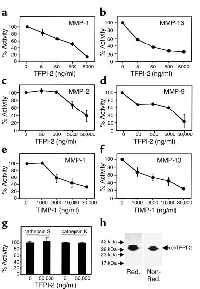
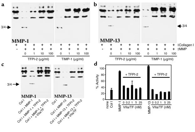
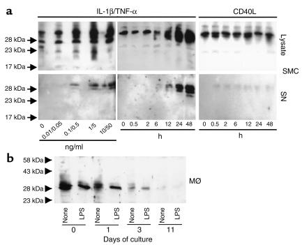
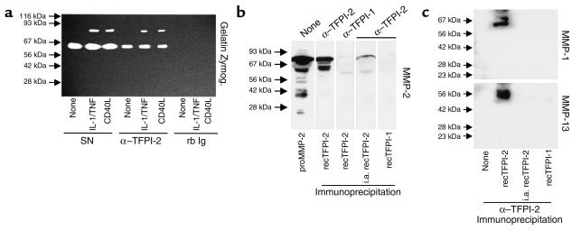
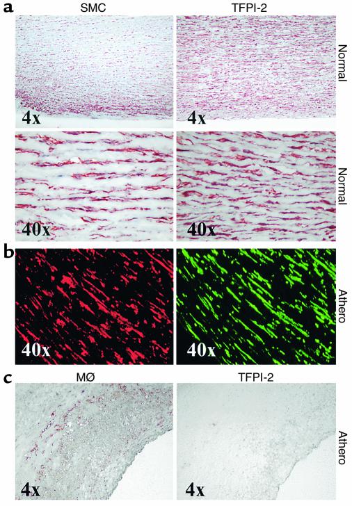
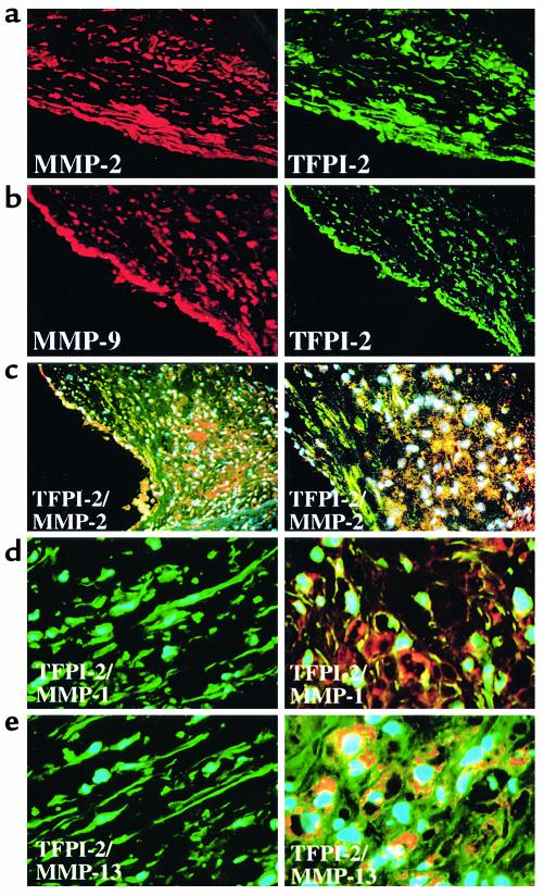
Degradation of ECM, particularly interstitial collagen, promotes plaque instability, rendering atheroma prone to rupture. Previous studies implicated matrix metalloproteinases (MMPs) in these processes, suggesting that dysregulated MMP activity, probably due to imbalance with endogenous inhibitors, promotes complications of atherosclerosis. We report here that the serine proteinase inhibitor tissue factor pathway inhibitor-2 (TFPI-2) can function as an MMP inhibitor. TFPI-2 diminished the ability of the interstitial collagenases MMP-1 and MMP-13 to degrade triple-helical collagen, the primary load-bearing molecule of the ECM within human atheroma. In addition, TFPI-2 also reduced the activity of the gelatinases MMP-2 and MMP-9. In contrast to the "classical" tissue inhibitors of MMPs (TIMPs), TFPI-2 expression in situ correlated inversely with MMP levels in human atheroma. TFPI-2 colocalized primarily with smooth muscle cells in the normal media as well as the plaque's fibrous cap. Conversely, the macrophage-enriched shoulder region, the prototypical site of matrix degradation and plaque rupture, stained only weakly for TFPI-2 but intensely for gelatinases and interstitial collagenases. Evidently, human mononuclear phagocytes, an abundant source of MMPs within human atheroma, lost their ability to express this inhibitor during differentiation in vitro. These findings establish a new, anti-inflammatory function of TFPI-2 of potential pathophysiological significance for human diseases, including atherosclerosis.


Figures

Figure 1
Figure 1
Human recombinant TFPI-2 (recTFPI-2) inhibits MMP activity. Human recombinant MMP-1 (a, e), MMP-13 (b, f), MMP-2 (c), MMP-9 (d), and cathepsin S or K (g) (all at 10 μg/ml) were incubated with the respective concentrations of TFPI-2 (ad, g) or TIMP-1 (e, f) (1 hour, 37°C), and the ability of the enzymes to process fluorophore-conjugated native (a, b, e, f) or peptide (c, d, g) substrate was measured as described. Percentage of activity was normalized to the activity obtained with the respective MMP lacking TFPI-2. Data shown represent mean ± SD and are representative of at least three independent experiments. (h) Human recombinant TFPI-2 (10 μg/lane), expressed and purified as described in Methods, was applied to 15% SDS-PAGE analysis under reducing (Red.) and nonreducing (Non-Red.) conditions. Proteins were visualized by silver staining. The positions of the molecular-weight markers are indicated on the left.
Figure 2
Figure 2
TFPI-2 inhibits processing of triple-helical type I collagen (Col I) by interstitial collagenases. Purified human triple-helical type I collagen (100 μg/ml) was incubated (24 hours, 25°C) with either MMP-1 (a) or MMP-13 (b) (both at 10 μg/ml) preincubated with the respective concentration of TFPI-2 or TIMP-1, or (c) incubated with MMP-1 and MMP-13 (both at 10 μg/ml) preincubated with TFPI-2 (100 μg/ml) complexed (45 minutes, 37°C) with VIIa/TF (20 nM). Reactions were applied to Western blot analysis with anti–type I collagen Ab. (d) Human recombinant MMP-1 or MMP-13 (both at 10 μg/ml) were incubated with TFPI-2 (100 μg/ml) complexed with the respective concentrations of VIIa/TF, and the subsequent ability of the MMPs to process native fluorogenic substrate was measured as described. Percentage of activity was normalized to the activity obtained with the respective MMP lacking TFPI-2. Data shown represent mean ± SD and are representative of at least three independent experiments.
Figure 3
Figure 3
Differential expression of TFPI-2 in cultured SMCs and MØs. (a) Human vascular SMCs were cultured in IT medium (24 hours) and either stimulated 24 hours with the respective concentrations of IL-1β/TNF-α (left), or stimulated for the indicated times with 10/50 ng/ml IL-1β/TNF-α (middle) or 10 μg/ml CD40L (right). (b) Mononuclear phagocytes (MØ) were cultured for either 0, 1, 3, or 11 days in RPMI-1640 medium supplemented with 5% human serum. Before (24 hours) stimulation with LPS (100 ng/ml), cells were cultured in serum-free medium. SMC and MØ culture lysates (30 μg/lane) and SMC supernatants (SN) were applied to Western blot analysis employing α–TFPI-2. The positions of the molecular-weight markers are indicated on the left. Comparable data were obtained employing cells from four different donors.
Figure 4
Figure 4
Anti–TFPI-2 coprecipitates MMP-1, MMP-2, MMP-9, and MMP-13. (a) Supernatants of unstimulated (none), IL-1β/TNF-α–stimulated (10/50 ng/ml), or CD40L-stimulated (10 μg/ml) human vascular SMCs were immunoprecipitated with anti–TFPI-2 (α–TFPI-2) or rabbit Ig (rb Ig), and the precipitates as well as the original culture supernatants (SN) were analyzed by SDS-PAGE zymography. Clear areas indicate gelatinolysis. Comparable data were obtained employing SMCs from three different donors. (b) Recombinant MMP-2 precursor as well as (c) active MMP-1 (top) and MMP-13 (bottom) (all 10 μg/ml) were incubated (1 hour, 37°C) with recombinant TFPI-2 (50 μg/ml). Reactions were immunoprecipitated with anti–TFPI-2 and analyzed by Western blotting for the respective MMP. For control purposes heat-inactivated recombinant TFPI-2 (i.a. recTFPI-2), recombinant TFPI-1 (50 μg/ml), or α–TFPI-1 was applied. The positions of the molecular-weight markers are indicated on the left.
Figure 5
Figure 5
SMCs and MØs differentially express TFPI-2 in human atheroma. Cryostat sections from nondiseased aortae (Normal) and atherosclerotic carotid atheroma (Athero) were stained for SMCs and MØs employing (a, b) anti–α-actin and (c) α-CD68, respectively (left panels). Localization of TFPI-2 (right panels) was shown by (a, c) light microscopy in adjacent sections, or (b) immunofluorescence double labeling for direct colocalization within the same section (α-actin staining for SMCs in red; TFPI-2 staining in green). Analysis of surgical specimens obtained from four different donors showed similar results.
Figure 6
Figure 6
TFPI-2 expression is diminished in the shoulder region of human atherosclerotic plaques. (a, b) Shown are single-wavelength filter photomicrographs (×10) of cryostat sections from atherosclerotic carotid atheroma applied to double-immunofluorescence labeling to colocalize (a) MMP-2 or (b) MMP-9 (both red) with TFPI-2 (green) within the SMC-enriched fibrous cap. (c) Triple-immunofluorescence labeling colocalized MMP-2 (red) with TFPI-2 (green) within the plaque’s shoulder region (left: ×4, right: ×10). Nuclei are stained in blue. High-magnification photomicrographs (×40) demonstrated differential expression of (d) MMP-1 or (e) MMP-13 (both red) with TFPI-2 (green) in SMC-enriched (left) or MØ-enriched (right) areas of human atheroma; nuclei are stained in blue. (f) Protein extracts from frozen tissue of nonatherosclerotic carotid arteries (Normal), as well as carotid plaques dichotomized into lesions displaying features associated with either stable (Fibrous) or vulnerable (Athero) plaques, were analyzed by Western blotting employing α–TIMP-1 (top), –TIMP-2 (middle), or –TFPI-2 (bottom). Analysis of five normal arteries, five fibrous and seven atheromatous surgical specimens of atheroma from different donors showed similar results.

Similar articles

Cited by

References

    1. Sprecher CA, Kisiel W, Mathewes S, Foster DC. Molecular cloning, expression, and partial characterization of a second human tissue-factor-pathway inhibitor. Proc Natl Acad Sci USA. 1994;91:3353–3357. - PMC - PubMed
    1. Petersen LC, et al. Inhibitory properties of a novel human Kunitz-type protease inhibitor homologous to tissue factor pathway inhibitor. Biochemistry. 1996;35:266–272. - PubMed
    1. Butzow R, Huhtala ML, Bohn H, Virtanen I, Seppala M. Purification and characterization of placental protein 5. Biochem Biophys Res Commun. 1988;150:483–490. - PubMed
    1. Iino M, Foster DC, Kisiel W. Quantification and characterization of human endothelial cell-derived tissue factor pathway inhibitor-2. Arterioscler Thromb Vasc Biol. 1998;18:40–46. - PubMed
    1. Rao CN, Mohanam S, Puppala A, Rao JS. Regulation of ProMMP-1 and ProMMP-3 activation by tissue factor pathway inhibitor-2/matrix-associated serine protease inhibitor. Biochem Biophys Res Commun. 1999;255:94–98. - PubMed

Publication types

MeSH terms