. 2001 May;107(9):1145-52.
doi: 10.1172/JCI12089.

Impaired IL-18 processing protects caspase-1-deficient mice from ischemic acute renal failure

Affiliations

Impaired IL-18 processing protects caspase-1-deficient mice from ischemic acute renal failure

V Y Melnikov et al. J Clin Invest. 2001 May.

Abstract

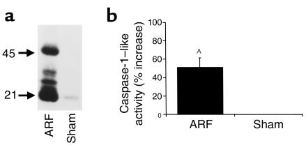
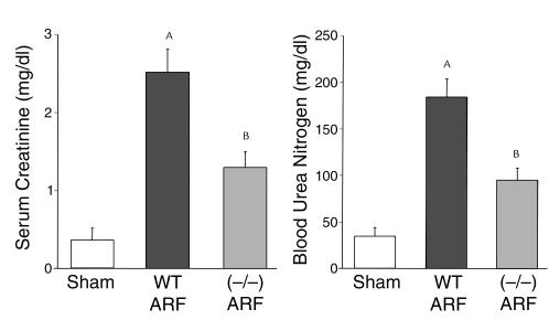
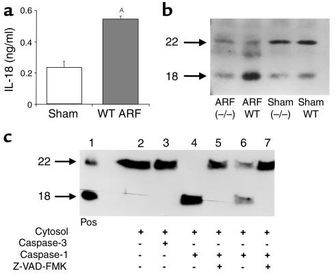
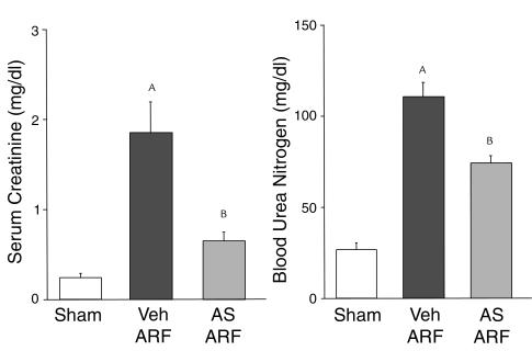
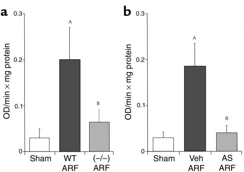
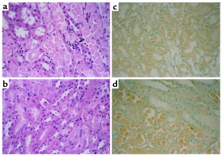
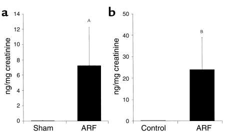
We sought to determine whether mice deficient in the proinflammatory caspase-1, which cleaves precursors of IL-1 beta and IL-18, were protected against ischemic acute renal failure (ARF). Caspase-1(-/-) mice developed less ischemic ARF as judged by renal function and renal histology. These animals had significantly reduced blood urea nitrogen and serum creatinine levels and a lower morphological tubular necrosis score than did wild-type mice with ischemic ARF. Since caspase-1 activates IL-18, lack of mature IL-18 might protect these caspase-1(-/-) mice from ARF. In wild-type animals, we found that ARF causes kidney IL-18 levels to more than double and induces the conversion of the IL-18 precursor to the mature form. This conversion is not observed in caspase-1(-/-) ARF mice or sham-operated controls. We then injected wild-type mice with IL-18-neutralizing antiserum before the ischemic insult and found a similar degree of protection from ARF as seen in caspase-1(-/-) mice. In addition, we observed a fivefold increase in myeloperoxidase activity in control mice with ARF, but no such increase in caspase-1(-/-) or IL-18 antiserum-treated mice. Finally, we confirmed histologically that caspase-1(-/-) mice show decreased neutrophil infiltration, indicating that the deleterious role of IL-18 in ischemic ARF may be due to increased neutrophil infiltration.


Figures

Figure 1
Figure 1
Caspase-1 protein expression (a) and activity (b) are increased in ischemic ARF. In wild-type mice, there was an increase in protein expression of both pro–caspase-1 (45 kDa) and active caspase-1 (21 kDa) in ischemic ARF compared with sham-operated controls (Sham). The immunoblot shown is representative of three separate experiments (a). Also, in wild-type mice, there was an increase in caspase-1–like activity in ischemic ARF compared with sham-operated controls (AP < 0.01 vs. sham, n = 7) (b).
Figure 2
Figure 2
Caspase-1–/– mice are protected against ischemic ARF. Caspase-1–/– mice developed less severe ARF, as determined by BUN and serum creatinine, compared with sham-operated wild-type (WT) mice. AP < 0.001 vs. sham; BP < 0.01 vs. WT ARF; n = 8.
Figure 3
Figure 3
IL-18 in ischemic ARF. (a) IL-18 protein, measured by the ECL assay, was increased in ischemic ARF in wild-type (WT) mice compared with sham-operated controls (sham). AP < 0.01 vs. sham, n = 6. (b) In sham-operated wild-type (WT) and caspase-1–/– mice, IL-18 was predominantly in the pro–IL-18 form (22 kDa). In wild-type mice with ischemic ARF (ARF WT), there was a conversion of the pro–IL-18 form (22 kDa) to the active form (18 kDa). This conversion of the pro–IL-18 to active IL-18 form was attenuated in caspase-1–/– mice with ischemic ARF (ARF–/–). The immunoblot shown is representative of three separate experiments. (c) In an in vitro experiment, cytosolic extracts from normal wild-type WT mouse kidney were incubated with purified caspases. Recombinant murine pro–IL-18 (22 kDa) and active IL-18 (18 kDa) were used as a positive control (Pos) (lane 1). In the cytosolic extract with no additions, IL-18 was present only in the precursor form (lane 2) and addition of purified caspase-3 (10 ng) had no effect (lane 3). Addition of purified caspase-1 (10 ng) completely cleaved IL-18 from the precursor to active form (lane 4) and caspase-1 (1 ng) partially cleaved IL-18 (lane 6). Prior incubation with the caspase inhibitor, Z-VAD-FMK, prevents the cleavage of pro–IL-18 to active IL-18 (lanes 5 and 7). These data demonstrate that caspase-1, but not caspase-3, cleaves IL-18 in the mouse kidney.
Figure 4
Figure 4
Mice treated with rabbit anti-murine IL-18–neutralizing antiserum are protected against ischemic ARF. Wild-type mice treated with anti–IL-18 antiserum (AS) developed less severe ischemic ARF compared with vehicle-treated (normal rabbit serum; Veh) mice. AP < 0.001 vs. sham, BP < 0.05 vs. ARF Veh; n = 6.
Figure 5
Figure 5
The increase in MPO activity during ischemic ARF is blocked in caspase-1–/– mice (a) and in mice treated with anti–IL-18 antiserum (b). In wild-type mice with ischemic ARF (WT ARF), there was an increase in MPO activity compared with sham-operated controls. In caspase-1–/– mice with ischemic ARF, MPO activity was normalized. AP < 0.001 vs. sham; BP < 0.01 vs. WT ARF; and NS vs. sham; n = 6 (a). In separate experiments, in wild-type mice treated with anti–IL-18 antiserum before ischemic ARF (AS ARF), MPO activity was normalized compared with vehicle-treated (Veh) animals. AP < 0.001 vs. sham; BP < 0.01 vs. Veh ARF; and NS vs. sham; n = 6 (b).
Figure 6
Figure 6
Renal histopathology (representative picture of at least three experiments). In wild-type mice with ischemic ARF, proximal tubules in the outer medulla show extensive damage including epithelial cell sloughing with focal denudation and numerous neutrophils (arrow) (a). In comparable sections from caspase-1–/– mice, tubules are largely intact with only focal sloughing of tubular cytoplasm and minimal loss of brush border. Neutrophils are inconspicuous in this case (b). Immunohistochemistry for IL-18 showed that there was cytoplasmic immunoreactivity in proximal tubule epithelium in sham-operated wild-type mice (c). There was increased IL-18 staining in necrotic proximal tubule epithelium in ischemic ARF in wild-type mice (d).
Figure 7
Figure 7
IL-18 was measured in the urine of mice with ARF by the ECL method that detects both the pro–IL-18 form and mature IL-18 form (a). IL-18 was also measured in the urine of human patients with ARF using a human IL-18 ELISA kit that detects the mature form of IL-18 (b). IL-18 levels were expressed per milligram of urinary creatinine to correct for differences in urine concentration. AP < 0.05 vs sham; BP = 0.01 vs controls; n = 4 for mouse and n = 5 for patients.

References

    1. Kaushal GP, Ueda N, Shah SV. Role of caspases (ICE/CED 3 proteases) in DNA damage and cell death in response to a mitochondrial inhibitor, antimycin A. Kidney Int. 1997;52:438–445. - PubMed
    1. Edelstein CL, Shi Y, Schrier RW. Role of caspases in hypoxia-induced necrosis of rat renal proximal tubules. J Am Soc Nephrol. 1999;10:1940–1949. - PubMed
    1. Daemen MARC, et al. Inhibition of apoptosis induced by ischemia-reperfusion prevents inflammation. J Clin Invest. 1999;104:541–549. - PMC - PubMed
    1. Fraser A, Evan G. A license to kill. Cell. 1996;85:781–784. - PubMed
    1. Dinarello CA. Biologic basis for interleukin-1 in disease. Blood. 1996;87:2095–2147. - PubMed

Publication types