. 2001 May;107(9):1183-92.
doi: 10.1172/JCI11774.

Heparin-binding defective lipoprotein lipase is unstable and causes abnormalities in lipid delivery to tissues

Affiliations

Heparin-binding defective lipoprotein lipase is unstable and causes abnormalities in lipid delivery to tissues

E P Lutz et al. J Clin Invest. 2001 May.

Abstract

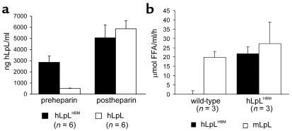
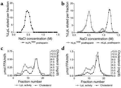
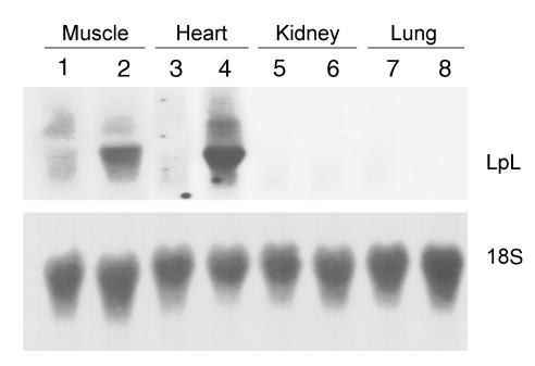
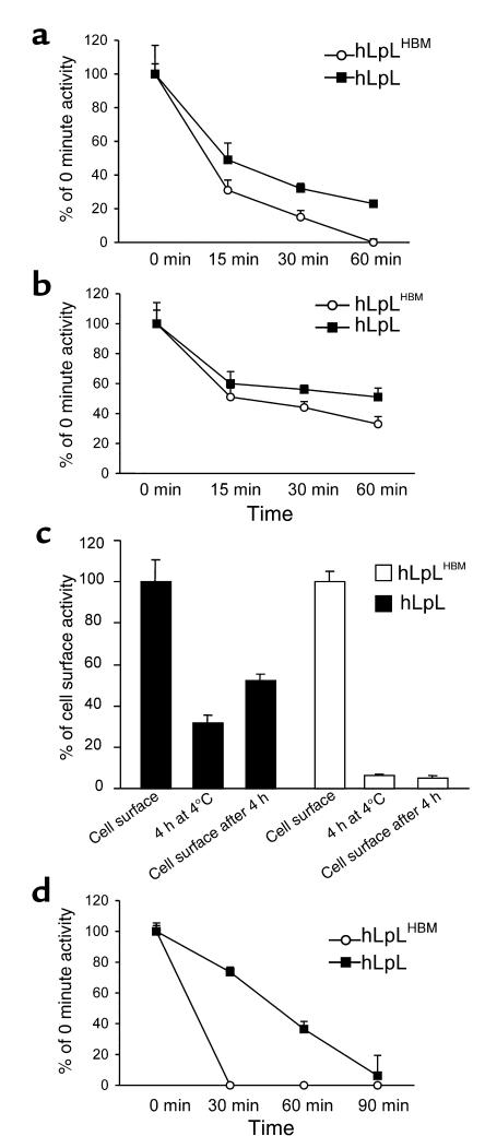
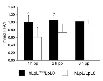
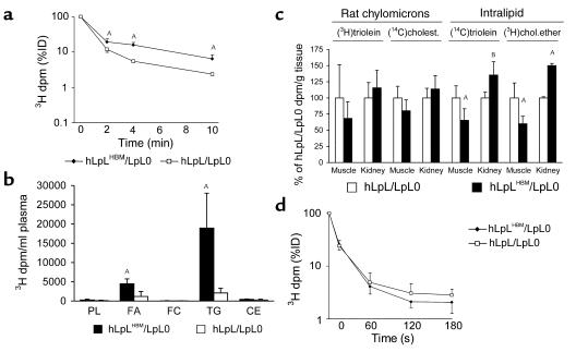
Lipoprotein lipase (LpL) binding to heparan sulfate proteoglycans (HSPGs) is hypothesized to stabilize the enzyme, localize LpL in specific capillary beds, and route lipoprotein lipids to the underlying tissues. To test these hypotheses in vivo, we created mice expressing a human LpL minigene (hLpL(HBM)) carrying a mutated heparin-binding site. Three basic amino acids in the carboxyl terminal region of LpL were mutated, yielding an active enzyme with reduced heparin binding. Mice expressing hLpL(HBM) accumulated inactive human LpL (hLpL) protein in preheparin blood. hLpL(HBM) rapidly lost activity during a 37 degrees C incubation, confirming a requirement for heparin binding to stabilize LPL: Nevertheless, expression of hLpL(HBM) prevented the neonatal demise of LpL knockout mice. On the LpL-deficient background hLpL(HBM) expression led to defective targeting of lipids to tissues. Compared with mice expressing native hLpL in the muscle, hLpL(HBM) transgenic mice had increased postprandial FFAs, decreased lipid uptake in muscle tissue, and increased lipid uptake in kidneys. Thus, heparin association is required for LpL stability and normal physiologic functions. These experiments confirm in vivo that association with HSPGs can provide a means to maintain proteins in their stable conformations and to anchor them at sites where their activity is required.


Figures

Figure 1
Figure 1
hLpL mass and activity of hLpLHBM transgenic mice. (a) hLpL mass in pre- and postheparin plasma of hLpLHBM and hLpL transgenic mice. Preheparin plasma was collected immediately before intravenous injection of 100 U heparin/kg. Five minutes later postheparin plasma was obtained. hLpL mass was determined by ELISA as described in Methods. (b) hLpLHBM and murine LpL (mLpL) activity in postheparin plasma of hLpLHBM transgenic mice and their wild-type littermates. A monoclonal antibody against hLpL (28) was utilized to distinguish between hLpLHBM and mLpL activity.
Figure 2
Figure 2
Heparin-affinity chromatography and gel filtration of mouse plasma. (a) Preheparin plasma from hLpLHBM transgenic mice was collected and applied to a heparin-affinity gel. LpL was eluted with a NaCl gradient and hLpL was measured by ELISA. (b) Postheparin plasma from hLpLHBM transgenic mice was chromatographed on a heparin-affinity gel. Human and murine LpL masses were determined by ELISA. The amounts of LpL are given as a percentage of eluted LpL per fraction. (c) Pooled postheparin plasma from hLpLHBM/LpL0 mice was gel filtered on an FPLC system with two Superose 6 columns in series. LpL activity and cholesterol were determined in each fraction. (d) Pooled postheparin plasma from hLpL/LpL0 mice was gel filtered and analyzed as described in c.
Figure 3
Figure 3
LpL RNA tissue levels of hLpLHBM and hLpL transgenic mice on the LpL knockout background. Total RNA of the indicated tissues of hLpLHBM/LpL0 and hLpL/LpL0 mice was separated on a 1% agarose gel, blotted to a nylon membrane, and hybridized with the ∼500 bp RsrII/HindIII fragment of the hLpL minigene. Lanes 1 and 2 show a 90-minute autoradiography of the blot while the lanes 3–8 show a 16-hour exposure. hLpLHBM RNA is in lanes 2, 4, 6, and 8; hLpL-RNA is in lanes 1, 3, 5, and 7. For control, 18S RNA was hybridized with a complementary oligonucleotide.
Figure 4
Figure 4
LpL stability. (a) Media from CHO cells expressing nonmutated hLpL (filled squares) and hLpLHBM (open circles) were assayed for LpL activity before and at the indicated time points during incubation at 37°C. The activity before the incubation is set as 100%. (b) 5 U heparin/ml were added to the cell culture media, then the experiment was performed as in a. (c) The stability of hLpLHBM (open bars) associated with the cell surface of CHO cells was compared with hLpL (filled bars). The initial cell-associated LpL activity was determined by incubating the cells with 20 U/ml of heparin for 30 minutes at 4°C. This was set as 100%. Then LpL activity in heparin-free media after a 4-hour incubation at 4°C with the cells is shown. The third bar indicates the activity released from the cell surface with heparin after the 4-hour incubation described before. (d) Stability of postheparin plasma from hLpLHBM/LpL0 was compared with hLpL/LpL0. The samples were assayed for LpL activity before incubation at 37°C and at the indicated time points. The initial activity was set to 100%.
Figure 5
Figure 5
Postprandial FFAs. After 8 hours of daytime fasting, a gavage with 100 μl corn oil was performed. Blood was withdrawn from the retro-orbital plexus and plasma was assayed for FFA at the indicated time points. AP < 0.03. pp, postprandial.
Figure 6
Figure 6
Rat chylomicron, Intralipid, and palmitate turnover studies. (a) Rat chylomicrons were labeled in vivo with (3H)triolein, collected as described in Methods, and injected into the tail vein of hLpLHBM/LpL0 (n = 5) and hLpL/LpL0 (n = 5) male mice. Blood was collected 2, 4, and 10 minutes after injection. Total plasma volume was calculated as 2.75% of body weight. Data are expressed as percentage of injected dose (ID). (b) A 100 μl aliquot of the plasma from the 10-minute time point was lipid extracted and separated by TLC. The bars show the 3H dpm per ml plasma for the indicated lipid. (PL, phospholipids; FA, fatty acids; FC, free cholesterol; CE, cholesterol esters). (c) Ten minutes after injection of labeled rat chylomicrons or Intralipid (injected into four hLpLHBM/LpL0 and four hLpL/LpL0 female mice), the mice were perfused with PBS and the indicated organs were taken out. Lipids were extracted with chloroform-methanol 2:1 (42) and counted. The data are expressed as percentage of hLpL/LpL0 dpm/g tissue. (d) (3H)palmitate was complexed to fatty acid–free BSA and injected into four hLpLHBM/LpL0 and four hLpL/LpL0 male mice. Mice were bled at the indicated time points. Data are expressed as percentage of injected dose. AP ≤ 0.02; BP ≤ 0.05. Cholest., cholesterol; chol.ether, cholesterol ether.
Figure 7
Figure 7
Histological examination of muscle from hLpLHMB transgenic mice. Periodic-acid Schiff–stained (PAS-stained) sections of quadriceps muscles of an 8-month-old female hLpLHBM/LpL2 mouse. In most areas completely normal histology was found (a). Only in small areas of hLpLHBM/LpL2 quadriceps some discrete, nonspecific myopathic alterations including more frequently centralized nuclei, a low number of in part atrophic muscle fibers, and some lipocytes in the endomysium could be found. Rarely a slight increase in glycogen storage could be detected (b). Compared with mice expressing high levels of hLpL, these findings are minimal. White arrow shows centralized nucleus, yellow arrow shows glycogen.

References

    1. Goldberg IJ. Lipoprotein lipase and lipolysis: central roles in lipoprotein metabolism and atherogenesis. J Lipid Res. 1996;37:693–707. - PubMed
    1. Iverius PH, Lindahl U, Egelrud T, Olivecrona T. Effects of heparin on lipoprotein lipase from bovine milk. J Biol Chem. 1972;247:6610–6616. - PubMed
    1. Shimada K, Gill PJ, Silbert JE, Douglas WH, Fanburg BL. Involvement of cell surface heparin sulfate in the binding of lipoprotein lipase to cultured bovine endothelial cells. J Clin Invest. 1981;68:995–1002. - PMC - PubMed
    1. Saxena U, Klein MG, Goldberg IJ. Identification and characterization of the endothelial cell surface lipoprotein lipase receptor. J Biol Chem. 1991;266:17516–17521. - PubMed
    1. Parthasarathy N, et al. Oligosaccharide sequences of endothelial cell surface heparan sulfate proteoglycan with affinity for lipoprotein lipase. J Biol Chem. 1994;269:22391–22396. - PubMed

Publication types

MeSH terms