. 2001 May 7;193(9):1027-34.
doi: 10.1084/jem.193.9.1027.

Hypoxia-inducible factor 1-dependent induction of intestinal trefoil factor protects barrier function during hypoxia

Affiliations

Hypoxia-inducible factor 1-dependent induction of intestinal trefoil factor protects barrier function during hypoxia

G T Furuta et al. J Exp Med. .

Abstract

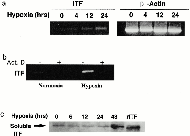
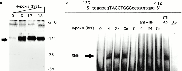
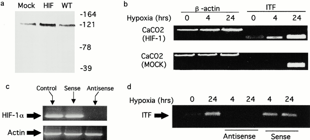
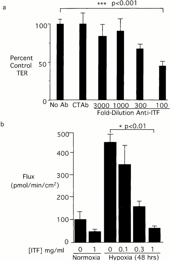
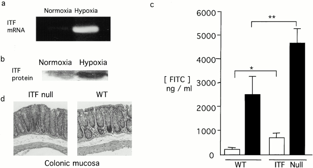
Mucosal organs such as the intestine are supported by a rich and complex underlying vasculature. For this reason, the intestine, and particularly barrier-protective epithelial cells, are susceptible to damage related to diminished blood flow and concomitant tissue hypoxia. We sought to identify compensatory mechanisms that protect epithelial barrier during episodes of intestinal hypoxia. Initial studies examining T84 colonic epithelial cells revealed that barrier function is uniquely resistant to changes elicited by hypoxia. A search for intestinal-specific, barrier-protective factors revealed that the human intestinal trefoil factor (ITF) gene promoter bears a previously unappreciated binding site for hypoxia-inducible factor (HIF)-1. Hypoxia resulted in parallel induction of ITF mRNA and protein. Electrophoretic mobility shift assay analysis using ITF-specific, HIF-1 consensus motifs resulted in a hypoxia-inducible DNA binding activity, and loading cells with antisense oligonucleotides directed against the alpha chain of HIF-1 resulted in a loss of ITF hypoxia inducibility. Moreover, addition of anti-ITF antibody resulted in a loss of barrier function in epithelial cells exposed to hypoxia, and the addition of recombinant human ITF to vascular endothelial cells partially protected endothelial cells from hypoxia-elicited barrier disruption. Extensions of these studies in vivo revealed prominent hypoxia-elicited increases in intestinal permeability in ITF null mice. HIF-1-dependent induction of ITF may provide an adaptive link for maintenance of barrier function during hypoxia.


Figures

Figure 1
Figure 1
ITF induction of hypoxia. Confluent Caco-2 epithelial monolayers were exposed to indicated periods of ambient hypoxia (pO2 = 20 Torr) or normoxia (pO2 = 147 Torr). In a, total RNA was isolated and examined for ITF transcript by RT-PCR. β-Actin transcript was used as an internal control. In b, Caco-2 cells were pretreated with vehicle or actinomycin D (Act. D) before 24 h of hypoxia or normoxia. Total RNA was isolated and examined for ITF mRNA. In c, soluble supernatants derived from monolayers exposed to indicated periods of hypoxia were examined for ITF content by SDS-PAGE and Western blot analysis.
Figure 2
Figure 2
Parallel induction of HIF-1 and ITF by hypoxia. Confluent Caco-2 epithelial monolayers were exposed to indicated periods of ambient hypoxia (pO2 = 20 Torr) or normoxia (pO2 = 147 Torr). In a, nuclear lysates derived from monolayers exposed to normoxia or hypoxia were examined for HIF-1α content by SDS-PAGE Western blot analysis. In b, interactions between HIF-1α and the HRE of the ITF promoter gene were examined by EMSA using synthetic oligonucleotides and nuclear lysates derived from cells exposed to normoxia, 4- or 24-h hypoxia, or CoCl2 (100 μM for 4 h). Specificity was determined with the use of anti–HIF-1α Ab, anti-CREB Ab (control), or excess (XS) unlabeled probed. Note diminution of signal in the presence of anti–HIF-1α, but not by anti-CREB Ab.
Figure 3
Figure 3
Induction of ITF is HIF-1α dependent. (a) Analysis of HIF-1α in transfected Caco-2 cells. Shown are mock transfected (Mock), cells overexpressing HIF-1α (HIF), and wild-type (WT) Caco-2 cells exposed to hypoxia and examined for HIF-1α expression by Western blot analysis. In b, ITF expression was analyzed by RT-PCR in HIF-1α transfectants and mock transfectants after exposure to indicated periods of hypoxia. In c, cells were loaded with antisense or sense oligonucleotides directed against HIF-1α as indicated and examined for HIF-1α mRNA. In d, Caco-2 cells pretreated with sense or antisense oligonucleotides as indicated were exposed to hypoxia or normoxia, and ITF mRNA expression was analyzed by RT-PCR.
Figure 4
Figure 4
Functional role of ITF peptide on barrier function. In a, Caco-2 cells were grown to electrical confluence on polycarbonate supports. Anti-ITF immune serum at indicated concentrations or control anti-CREB (1:100) was added to the apical well of the support. Cells were exposed to indicated periods of ambient hypoxia (pO2 = 20 Torr) or normoxia. Transepithelial electrical resistance was measured at 48 h and indicated as data normalized to untreated control monolayers. 1:100 anti-ITF versus no Ab; ***P < 0.001. (b) The influence of recombinant ITF protein on permeability changes elicited in human MVEC exposure to hypoxia. Endothelia were grown to confluence on polycarbonate supports. Recombinant human ITF at indicated concentrations were applied to the apical well, and cells were exposed to indicated periods of ambient hypoxia (pO2 = 20 Torr) or normoxia. Flux of 40-kD FITC-labeled dextran was used to assess endothelial paracellular permeability. 1 mg/ml hITF versus no recombinant protein; *P < 0.001.
Figure 5
Figure 5
Role of ITF in vivo. ITF null or wild-type Bl6129F1 mice were gavaged with 60 mg/100 g body weight of FITC-dextran and exposed to 4-h ambient hypoxia (8% O2) or ambient room air. Mice were killed, colonic tissue harvested for RNA (a), Western blot analysis (b), histologic analysis original magnification: ×10 (d), and serum FITC-dextran was quantified as a measure of intestinal permeability (c). *P < 0.05; **P < 0.01.

References

    1. Madara J.L. Regulation of the movement of solutes across tight junctions. Annu. Rev. Physiol. 1998;60:143–159. - PubMed
    1. Taylor C.T., Colgan S.P. Therapeutic targets for hypoxia-elicited pathways. Pharmacol. Res. 1999;16:1498–1505. - PubMed
    1. Semenza G.L. Perspectives on oxygen sensing. Cell. 1999;98:281–284. - PubMed
    1. Semenza G.L. Hypoxia-inducible factor 1master regulator of O2 homeostasis. Curr. Opin. Genet. Dev. 1998;8:588–594. - PubMed
    1. Wang G.L., Semenza G.L. Purification and characterization of hypoxia-inducible factor 1. J. Biol. Chem. 1995;270:1230–1237. - PubMed

Publication types

MeSH terms