. 2001 May 7;193(9):1035-44.
doi: 10.1084/jem.193.9.1035.

The adenylate cyclase toxin of Bordetella pertussis binds to target cells via the alpha(M)beta(2) integrin (CD11b/CD18)

Affiliations

The adenylate cyclase toxin of Bordetella pertussis binds to target cells via the alpha(M)beta(2) integrin (CD11b/CD18)

P Guermonprez et al. J Exp Med. .

Abstract

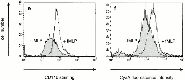
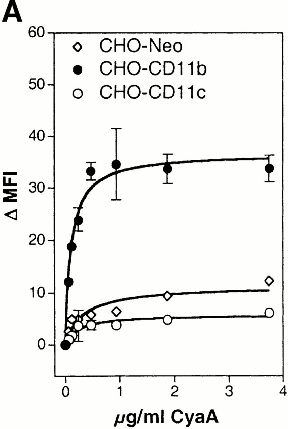
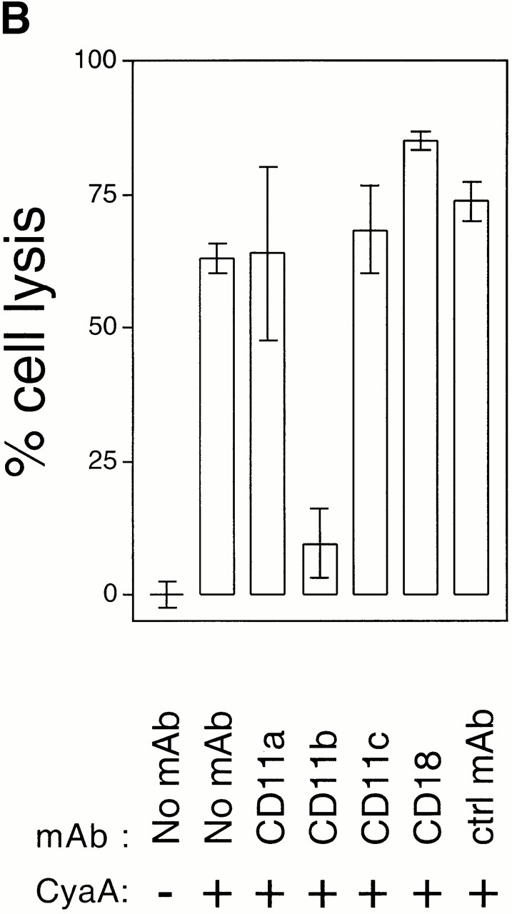
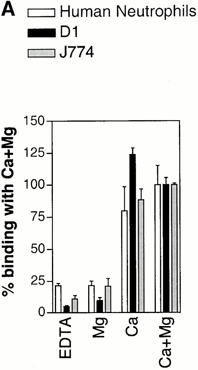
The adenylate cyclase toxin (CyaA) of Bordetella pertussis is a major virulence factor required for the early phases of lung colonization. It can invade eukaryotic cells where, upon activation by endogenous calmodulin, it catalyzes the formation of unregulated cAMP levels. CyaA intoxication leads to evident toxic effects on macrophages and neutrophils. Here, we demonstrate that CyaA uses the alpha(M)beta(2) integrin (CD11b/CD18) as a cell receptor. Indeed, the saturable binding of CyaA to the surface of various hematopoietic cell lines correlated with the presence of the alpha(M)beta(2) integrin on these cells. Moreover, binding of CyaA to various murine cell lines and human neutrophils was specifically blocked by anti-CD11b monoclonal antibodies. The increase of intracellular cAMP level and cell death triggered by CyaA intoxication was also specifically blocked by anti-CD11b monoclonal antibodies. In addition, CyaA bound efficiently and triggered intracellular cAMP increase and cell death in Chinese hamster ovary cells transfected with alpha(M)beta(2) (CD11b/CD18) but not in cells transfected with the vector alone or with the alpha(X)beta(2) (CD11c/CD18) integrin. Thus, the cellular distribution of CD11b, mostly on neutrophils, macrophages, and dendritic and natural killer cells, supports a role for CyaA in disrupting the early, innate antibacterial immune response.


Figures

Figure 1
Figure 1
Saturable binding of CyaA to different cell lines correlates with their CD11b expression. (A) CyaA binding at the surface of dendritic cells (D1), macrophages (J774A.1), B cells (LB27.4), and T cells (EL4) was performed at 37°C for 20 min. Surface-bound CyaA was detected by flow cytometry using biotinylated anti-CyaA polyclonal Abs and streptavidin-PE. Binding is expressed as ΔMFI = (MFI value of cells incubated with CyaA) − (MFI of cells without CyaA). (B–E) Cell-surface expression of β2 integrins on D1, J774A.1, LB27.4, and EL4 cells. CD11a (B), CD11b (C), CD11c (D), and CD18 (E) expression was determined by flow cytometry using specific mAbs coupled to PE. Integrin expression is expressed as ΔMFI = (MFI value of cells stained with specific mAb) − (MFI of cells stained with an isotype control mAb).
Figure 1
Figure 1
Saturable binding of CyaA to different cell lines correlates with their CD11b expression. (A) CyaA binding at the surface of dendritic cells (D1), macrophages (J774A.1), B cells (LB27.4), and T cells (EL4) was performed at 37°C for 20 min. Surface-bound CyaA was detected by flow cytometry using biotinylated anti-CyaA polyclonal Abs and streptavidin-PE. Binding is expressed as ΔMFI = (MFI value of cells incubated with CyaA) − (MFI of cells without CyaA). (B–E) Cell-surface expression of β2 integrins on D1, J774A.1, LB27.4, and EL4 cells. CD11a (B), CD11b (C), CD11c (D), and CD18 (E) expression was determined by flow cytometry using specific mAbs coupled to PE. Integrin expression is expressed as ΔMFI = (MFI value of cells stained with specific mAb) − (MFI of cells stained with an isotype control mAb).
Figure 2
Figure 2
CyaA binding to murine cell lines is blocked by anti-CD11b mAb. Cells were preincubated at 4°C for 15 min with or without 20 μg/ml of specific mAbs and then incubated at 4°C for 20 min with 5 μg/ml CyaA and 10 μg/ml of corresponding mAbs. Surface-bound CyaA was detected with biotinylated anti-CyaA polyclonal Abs, revealed by streptavidin-PE, and analyzed by flow cytometry on living cells. (a–c) Fluorescence histograms of D1 dendritic cells were preincubated with medium alone (a), with M1/70 anti-CD11b mAb (b), or with an isotype-matched rat IgG2b (c) and then incubated with (gray) or without (white) CyaA in the presence or absence of mAbs. The cell number is plotted against the log of PE fluorescence. (d–f) D1 dendritic cells (d), FSDCs (e), or J774A.1 macrophages (f) were preincubated with medium alone (○) or with M1/70 anti-CD11b mAb (•), and then incubated with CyaA with or without M1/70 anti-CD11b mAb. Bmax was determined by fitting experimental points to ΔMFI = B max × [CyaA]/(K d + [CyaA]). Binding of CyaA is plotted as a percentage of Bmax plotted against CyaA concentration. (g–i) Effect of specific mAbs on the binding of a fixed dose of CyaA. D1 (g), FSDCs (h), or J774A.1 (i) cells were preincubated with or without specific mAbs (anti-CD11a, 2D7, anti-CD11b, M1/70 or 5C6, anti-CD11c, HL3, anti-CD18, C17/16, control A95-1), and incubated with CyaA at the fixed concentration of 5 μg/ml. Values of ΔMFI obtained for CyaA binding on cells treated with specific mAbs were normalized as a percentage of the ΔMFI values obtained for CyaA binding without mAb.
Figure 2
Figure 2
CyaA binding to murine cell lines is blocked by anti-CD11b mAb. Cells were preincubated at 4°C for 15 min with or without 20 μg/ml of specific mAbs and then incubated at 4°C for 20 min with 5 μg/ml CyaA and 10 μg/ml of corresponding mAbs. Surface-bound CyaA was detected with biotinylated anti-CyaA polyclonal Abs, revealed by streptavidin-PE, and analyzed by flow cytometry on living cells. (a–c) Fluorescence histograms of D1 dendritic cells were preincubated with medium alone (a), with M1/70 anti-CD11b mAb (b), or with an isotype-matched rat IgG2b (c) and then incubated with (gray) or without (white) CyaA in the presence or absence of mAbs. The cell number is plotted against the log of PE fluorescence. (d–f) D1 dendritic cells (d), FSDCs (e), or J774A.1 macrophages (f) were preincubated with medium alone (○) or with M1/70 anti-CD11b mAb (•), and then incubated with CyaA with or without M1/70 anti-CD11b mAb. Bmax was determined by fitting experimental points to ΔMFI = B max × [CyaA]/(K d + [CyaA]). Binding of CyaA is plotted as a percentage of Bmax plotted against CyaA concentration. (g–i) Effect of specific mAbs on the binding of a fixed dose of CyaA. D1 (g), FSDCs (h), or J774A.1 (i) cells were preincubated with or without specific mAbs (anti-CD11a, 2D7, anti-CD11b, M1/70 or 5C6, anti-CD11c, HL3, anti-CD18, C17/16, control A95-1), and incubated with CyaA at the fixed concentration of 5 μg/ml. Values of ΔMFI obtained for CyaA binding on cells treated with specific mAbs were normalized as a percentage of the ΔMFI values obtained for CyaA binding without mAb.
Figure 3
Figure 3
CyaA binding to human neutrophils is blocked by anti-CD11b and anti-CD18 mAbs and is correlated to CD11b surface levels. (a–c) Fluorescence histograms of freshly purified neutrophils preincubated with medium alone (a), the anti-CD11b mAb 44 (b), or an isotype-matched control mouse mAb (c), then incubated with (gray) or without (white) biotinylated detoxified CyaA (5 μg/ml), and revealed by streptavidin-PE. Cell number is plotted against log of PE fluorescence. (d) Effect of specific mAbs on CyaA binding to neutrophils (anti-CD11b, 44 or M1/70, anti-CD18, TS/18, control mouse IgG2a, control rat IgG2b, A95-1). Freshly purified neutrophils were preincubated with or without specific mAbs and incubated with biotinylated detoxified CyaA. Values of ΔMFI obtained for CyaA binding on cells treated with specific mAbs were normalized as a percentage of the ΔMFI values obtained for CyaA binding without mAb. (e and f) Neutrophil activation triggers the increase of both CD11b surface level and CyaA binding. Fluorescence histograms of freshly purified neutrophils that were pretreated or not with 2 μM fMLP for 30 min at 37°C and then were stained with the anti-CD11b mAb (e) or 15 μg/ml biotinylated detoxified CyaA (f).
Figure 3
Figure 3
CyaA binding to human neutrophils is blocked by anti-CD11b and anti-CD18 mAbs and is correlated to CD11b surface levels. (a–c) Fluorescence histograms of freshly purified neutrophils preincubated with medium alone (a), the anti-CD11b mAb 44 (b), or an isotype-matched control mouse mAb (c), then incubated with (gray) or without (white) biotinylated detoxified CyaA (5 μg/ml), and revealed by streptavidin-PE. Cell number is plotted against log of PE fluorescence. (d) Effect of specific mAbs on CyaA binding to neutrophils (anti-CD11b, 44 or M1/70, anti-CD18, TS/18, control mouse IgG2a, control rat IgG2b, A95-1). Freshly purified neutrophils were preincubated with or without specific mAbs and incubated with biotinylated detoxified CyaA. Values of ΔMFI obtained for CyaA binding on cells treated with specific mAbs were normalized as a percentage of the ΔMFI values obtained for CyaA binding without mAb. (e and f) Neutrophil activation triggers the increase of both CD11b surface level and CyaA binding. Fluorescence histograms of freshly purified neutrophils that were pretreated or not with 2 μM fMLP for 30 min at 37°C and then were stained with the anti-CD11b mAb (e) or 15 μg/ml biotinylated detoxified CyaA (f).
Figure 5
Figure 5
CHO cells bind CyaA and become sensitive to CyaA when transfected with CD11b, but not with CD11c. (A and B) CyaA binding at the surface of CHO transfectants. CHO cells transfected with human CD11b/CD18, human CD11c/CD18, or mock-transfected were incubated with various doses of CyaA for 20 min at 37°C (A) or 4°C (B). Surface-bound CyaA was detected with biotinylated anti-CyaA polyclonal Abs, revealed by streptavidin-PE, and detected by flow cytometry on living cells. Binding is expressed as ΔMFI = (MFI value of cells incubated with CyaA) − (MFI of cells without CyaA). (C) Intracellular cAMP accumulation in CHO transfectants. CHO cells transfected with human CD11b/CD18, human CD11c/CD18, or mock-transfected were incubated with or without CyaA for 20 min at 37°C. Intracellular cAMP contents were determined as described in Materials and Methods. (D) Cell lysis in CHO transfectants. CHO cells transfected with human CD11b/CD18, human CD11c/CD18, or mock-transfected were incubated with 5 μg/ml CyaA for 4 h at 37°C. Cell lysis was determined by LDH release using the Cytotox 96™ assay.
Figure 5
Figure 5
CHO cells bind CyaA and become sensitive to CyaA when transfected with CD11b, but not with CD11c. (A and B) CyaA binding at the surface of CHO transfectants. CHO cells transfected with human CD11b/CD18, human CD11c/CD18, or mock-transfected were incubated with various doses of CyaA for 20 min at 37°C (A) or 4°C (B). Surface-bound CyaA was detected with biotinylated anti-CyaA polyclonal Abs, revealed by streptavidin-PE, and detected by flow cytometry on living cells. Binding is expressed as ΔMFI = (MFI value of cells incubated with CyaA) − (MFI of cells without CyaA). (C) Intracellular cAMP accumulation in CHO transfectants. CHO cells transfected with human CD11b/CD18, human CD11c/CD18, or mock-transfected were incubated with or without CyaA for 20 min at 37°C. Intracellular cAMP contents were determined as described in Materials and Methods. (D) Cell lysis in CHO transfectants. CHO cells transfected with human CD11b/CD18, human CD11c/CD18, or mock-transfected were incubated with 5 μg/ml CyaA for 4 h at 37°C. Cell lysis was determined by LDH release using the Cytotox 96™ assay.
Figure 4
Figure 4
Intracellular cAMP increase and cell death mediated by CyaA are specifically blocked by an anti-CD11b mAb in J774A.1 cells. (A) Effect of specific mAbs on intracellular cAMP accumulation. J774A.1 cells were preincubated at 4°C for 1 h with or without 10 μg/ml of specific mAbs (anti-CD11b, M1/70 or anti-CD18, C17/16) and then incubated at 37°C for 20 min with 5 μg/ml CyaA and with 10 μg/ml mAbs if present during the preincubation. Intracellular cAMP contents were determined as described in Materials and Methods. (B) Effect of specific mAbs on CyaA-mediated cell death. J774A.1 cells were preincubated at 4°C for 1 h with medium alone or with 10 μg/ml of specific mAbs (anti-CD11a 2D7, anti-CD11b M1/70, anti-CD11c HL3, anti-CD18 C71/16, control 2.4G2). Then, they were incubated at 37°C for 2 h with 0.5 μg/ml CyaA and with 10 μg/ml of specific mAbs when added during the preincubation. Cell lysis was determined by LDH release using the Cytotox 96™ assay.
Figure 4
Figure 4
Intracellular cAMP increase and cell death mediated by CyaA are specifically blocked by an anti-CD11b mAb in J774A.1 cells. (A) Effect of specific mAbs on intracellular cAMP accumulation. J774A.1 cells were preincubated at 4°C for 1 h with or without 10 μg/ml of specific mAbs (anti-CD11b, M1/70 or anti-CD18, C17/16) and then incubated at 37°C for 20 min with 5 μg/ml CyaA and with 10 μg/ml mAbs if present during the preincubation. Intracellular cAMP contents were determined as described in Materials and Methods. (B) Effect of specific mAbs on CyaA-mediated cell death. J774A.1 cells were preincubated at 4°C for 1 h with medium alone or with 10 μg/ml of specific mAbs (anti-CD11a 2D7, anti-CD11b M1/70, anti-CD11c HL3, anti-CD18 C71/16, control 2.4G2). Then, they were incubated at 37°C for 2 h with 0.5 μg/ml CyaA and with 10 μg/ml of specific mAbs when added during the preincubation. Cell lysis was determined by LDH release using the Cytotox 96™ assay.
Figure 6
Figure 6
Ca2+ is necessary and sufficient for CyaA binding to CD11b+ cells. (A) D1 or J774 cells, or freshly purified neutrophils were washed twice with Tris-buffered saline and then incubated with Tris-buffered saline supplemented with either 5 mM EDTA (EDTA), 2 mM Mg2+ and 100 μM EGTA (Mg), 2 mM Ca2+ (Ca), or 2 mM Ca2+ and 2 mM Mg2+ (Ca + Mg). After the addition of 10 μg/ml biotinylated detoxified CyaA, all samples were incubated for 20 min at 4°C. Bound CyaA was detected by streptavidin-PE as described above, and results are expressed as a percentage of CyaA binding in the presence of 2 mM Ca2+ and 2 mM Mg2+. (B) D1 cells were incubated in the presence of DMEM alone, DMEM and 5 mM EDTA, or DMEM and 5 mM EGTA, and stained with biotinylated detoxified CyaA as above.
Figure 6
Figure 6
Ca2+ is necessary and sufficient for CyaA binding to CD11b+ cells. (A) D1 or J774 cells, or freshly purified neutrophils were washed twice with Tris-buffered saline and then incubated with Tris-buffered saline supplemented with either 5 mM EDTA (EDTA), 2 mM Mg2+ and 100 μM EGTA (Mg), 2 mM Ca2+ (Ca), or 2 mM Ca2+ and 2 mM Mg2+ (Ca + Mg). After the addition of 10 μg/ml biotinylated detoxified CyaA, all samples were incubated for 20 min at 4°C. Bound CyaA was detected by streptavidin-PE as described above, and results are expressed as a percentage of CyaA binding in the presence of 2 mM Ca2+ and 2 mM Mg2+. (B) D1 cells were incubated in the presence of DMEM alone, DMEM and 5 mM EDTA, or DMEM and 5 mM EGTA, and stained with biotinylated detoxified CyaA as above.

References

    1. Weiss A.A., Hewlett E.L., Myers G.A., Falkow S. Pertussis toxin and extracytoplasmic adenylate cyclase as virulence factors of Bordetella pertussis . J. Infect. Dis. 1984;150:219–222. - PubMed
    1. Weiss A.A., Goodwin M.S. Lethal infection by Bordetella pertussis mutants in the infant mouse model. Infect. Immun. 1989;57:3757–3764. - PMC - PubMed
    1. Gross M.K., Au D.C., Smith A.L., Storm D.R. Targeted mutations that ablate either the adenylate cyclase or hemolysin function of the bifunctional cyaA toxin of Bordetella pertussis abolish virulence. Proc. Natl. Acad. Sci. USA. 1992;89:4898–4902. - PMC - PubMed
    1. Khelef N., Sakamoto H., Guiso N. Both adenylate cyclase and hemolytic activities are required by Bordetella pertussis to initiate infection. Microb. Pathog. 1992;12:227–235. - PubMed
    1. Khelef N., Bachelet C.M., Vargaftig B.B., Guiso N. Characterization of murine lung inflammation after infection with parental Bordetella pertussis and mutants deficient in adhesins or toxins. Infect. Immun. 1994;62:2893–2900. - PMC - PubMed

Publication types

MeSH terms