. 2001 May 22;98(11):6390-5.
doi: 10.1073/pnas.101564798. Epub 2001 May 8.

Leptin induces vascular permeability and synergistically stimulates angiogenesis with FGF-2 and VEGF

Affiliations

Leptin induces vascular permeability and synergistically stimulates angiogenesis with FGF-2 and VEGF

R Cao et al. Proc Natl Acad Sci U S A. .

Abstract

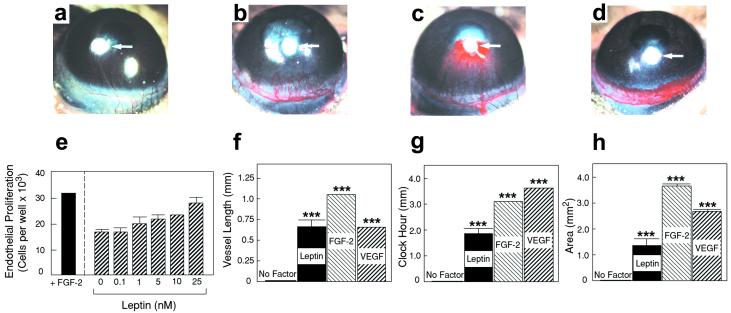
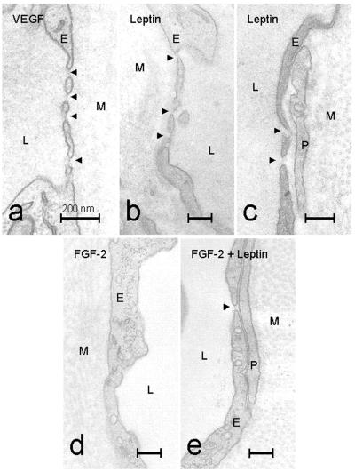
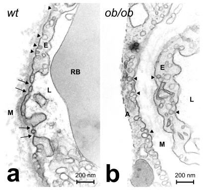
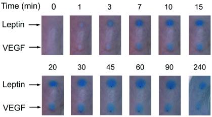
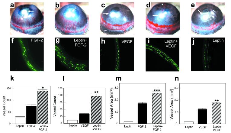
Most endocrine hormones are produced in tissues and organs with permeable microvessels that may provide an excess of hormones to be transported by the blood circulation to the distal target organ. Here, we investigate whether leptin, an endocrine hormone, induces the formation of vascular fenestrations and permeability, and we characterize its angiogenic property in the presence of other angiogenic factors. We provide evidence that leptin-induced new blood vessels are fenestrated. Under physiological conditions, capillary fenestrations are found in the leptin-producing adipose tissue in lean mice. In contrast, no vascular fenestrations were detected in the adipose tissue of leptin-deficient ob/ob mice. Thus, leptin plays a critical role in the maintenance and regulation of vascular fenestrations in the adipose tissue. Leptin induces a rapid vascular permeability response when administrated intradermally. Further, leptin synergistically stimulates angiogenesis with fibroblast growth factor (FGF)-2 and vascular endothelial growth factor (VEGF), the two most potent and commonly expressed angiogenic factors. These findings demonstrate that leptin has another new function-the increase of vascular permeability.


Figures

Figure 1
Figure 1
Endothelial cell growth and corneal angiogenic responses induced by leptin, FGF-2, and VEGF. (e) BCE cells were incubated with various concentrations of leptin or 1 ng/ml FGF-2. After 72 h, cells were counted. Values represent means (±SEM) of triplicate of each sample. Statistically significant values were obtained at concentrations above 5 nM (P = 0.085 at 5 nM, P = 0.0014 at 10 nM, and P = 0.027 at 25 nM) (a) A cornea of the control group of mice implanted with sucrose aluminum sulfate and Hydron (without growth factors). Pellets containing sucrose aluminum sulfate, Hydron, and 160 ng of leptin (b), 80 ng of FGF-2 (c), or 160 ng of VEGF (d) were implanted into corneal micropockets of mice. Corneas were photographed with a stereomicroscope on day 5 after pellet implantation; positions of implanted pellets are indicated by white arrows. Maximal vessel length (f), clock-hours of circumferential neovascularization (g), and area of neovascularization (h) are presented as mean determinants (±SEM) of 9 or 10 corneas in each group. P values were calculated according to a standard two-tailed Student's t test. ***, P < 0.001.
Figure 2
Figure 2
Thin parts of the walls of microvessels growing into the mouse cornea after stimulation with VEGF (a), leptin (b and c), FGF-2 (d) and FGF-2 + leptin (e). Arrowheads mark endothelial fenestrations. L, capillary lumen; M, collagenous matrix of the cornea; P, perivascular cell; E, endothelial cell. (Bars = 0.2 μm.)
Figure 3
Figure 3
Details of capillary structures in adipose tissue from (a) wild-type (wt) and (b) ob/ob mice. At least 200 capillary sections were examined in each group. Black arrows point to endothelial fenestrations. Arrowheads mark caveolae. A, adipocyte; L, capillary lumen; RB, red blood cell; E, endothelial cell; M, collagenous matrix of the cornea. (Bars = 0.2 μm.)
Figure 4
Figure 4
Evans blue was injected intravenously into the tail veins of BALB/c mice. After 5 min, 50 ng of leptin or VEGF was administered intradermally in 20 μl of PBS. The same amounts of FGF-2 and BSA were used as negative controls. The extravasation of Evans blue was recorded with a digital camera system at various time points (min).
Figure 5
Figure 5
(ae) Approximately 80 ng of leptin (e), 40 ng of FGF-2 (a), 80 ng of VEGF (c), and the same amounts of leptin plus FGF-2 (b) and leptin plus VEGF (d) were implanted into each corneal micropocket of mice. Corneas were photographed by a stereomicroscope on day 5 after growth factor implantation; positions of implanted pellets are indicated by white arrows in ae. The area of neovascularization (m and n) was calculated as described (28, 29) and presented as mean determinants (±SEM) of 10–12 corneas in each group. (fj) Corneal sections were incubated with an anti-CD31 antibody and stained with an FITC-conjugated secondary antibody. Immunohistological sections of the leptin-implanted cornea (j), FGF-2-implanted cornea (f), VEGF-implanted cornea (h), leptin plus FGF-2-implanted cornea (g), and leptin plus VEGF-implanted cornea (i). Corneal microvessels are revealed in green color. Vessel counts (k and l) are presented as mean determinants (±SEM) of 6–8 corneas in each group. *, P < 0.05; **, P < 0.01; ***, P < 0.001.

References

    1. Sierra-Honigmann M R, Nath A K, Murakami C, Garcia-Cardena G, Papapetropoulos A, Sessa W C, Madge L A, Schechner J S, Schwabb M B, Polverini P J, Flores-Riveros J R. Science. 1998;281:1683–1686. - PubMed
    1. Bouloumie A, Drexler H C, Lafontan M, Busse R. Circ Res. 1998;83:1059–1066. - PubMed
    1. Trayhurn P, Hoggard N, Mercer J G, Rayner D V. Int J Obes Relat Metab Disord. 1999;1:22–28. - PubMed
    1. Esser S, Wolburg K, Wolburg H, Breier G, Kurzchalia T, Risau W. J Cell Biol. 1998;140:947–959. - PMC - PubMed
    1. Roberts W G, Palade G E. J Cell Sci. 1995;108:2369–2379. - PubMed

Publication types

MeSH terms

LinkOut - more resources