. 2001 May 22;98(11):6144-9.
doi: 10.1073/pnas.101596998. Epub 2001 May 8.

Drosophila phosphoinositide-dependent kinase-1 regulates apoptosis and growth via the phosphoinositide 3-kinase-dependent signaling pathway

Affiliations

Drosophila phosphoinositide-dependent kinase-1 regulates apoptosis and growth via the phosphoinositide 3-kinase-dependent signaling pathway

K S Cho et al. Proc Natl Acad Sci U S A. .

Abstract

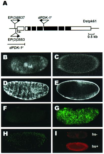
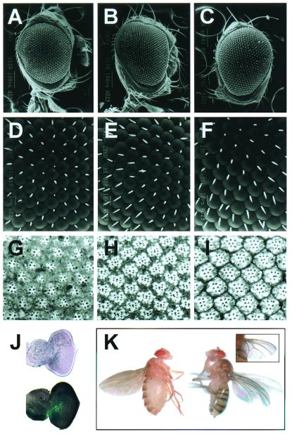
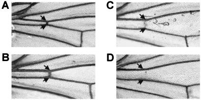
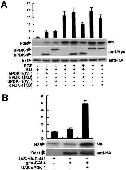
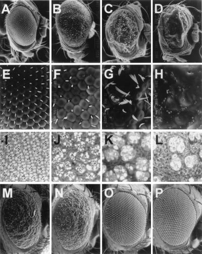
Phosphoinositide-dependent kinase-1 (PDK-1) is a central mediator of the cell signaling between phosphoinositide 3-kinase (PI3K) and various intracellular serine/threonine kinases including Akt/protein kinase B (PKB), p70 S6 kinases, and protein kinase C. Recent studies with cell transfection experiments have implied that PDK-1 may be involved in various cell functions including cell growth and apoptosis. However, despite its pivotal role in cellular signalings, the in vivo functions of PDK-1 in a multicellular system have rarely been investigated. Here, we have isolated Drosophila PDK-1 (dPDK-1) mutants and characterized the in vivo roles of the kinase. Drosophila deficient in the dPDK-1 gene exhibited lethality and an apoptotic phenotype in the embryonic stage. Conversely, overexpression of dPDK-1 increased cell and organ size in a Drosophila PI3K-dependent manner. dPDK-1 not only could activate Drosophila Akt/PKB (Dakt1), but also substitute the in vivo functions of its mammalian ortholog to activate Akt/PKB. This functional interaction between dPDK-1 and Dakt1 was further confirmed through genetic analyses in Drosophila. On the other hand, cAMP-dependent protein kinase, which has been proposed as a possible target of dPDK-1, did not interact with dPDK-1. In conclusion, our findings provide direct evidence that dPDK-1 regulates cell growth and apoptosis during Drosophila development via the PI3K-dependent signaling pathway and demonstrate our Drosophila system to be a powerful tool for elucidating the in vivo functions and targets of PDK-1.


Figures

Figure 1
Figure 1
Characterization of dPDK-1 mutants. (A) The insertion sites of the P-element in dPDK-1 mutants. The triangle represents the P-element. Two EP lines, EP(3)837 and EP(3)3553, have P-elements in the 5′ flanking region of dPDK-1 gene, and dPDK-11 has the P-element in the fourth intron. dPDK-12 was generated by an imprecise excision of P-element from EP(3)837. The deletion site of dPDK-12 is presented by a straight line, which includes the first exon of the kinase. (B) A heterozygous embryo of dPDK-11 (dPDK-11/TM3, Act-GFP, Ser1) at stage 16. (D and F) Wild-type embryos at stage 16. (C, E, and G) dPDK-11 homozygous embryos (dPDK-11/dPDK-11) at stage 16. (B and C) Fluorescent confocal views of heterozygous (B) and homozygous (C) embryos. GFP was expressed only in the heterozygous embryo (B) in the midgut and salivary glands. We could first detect the expression of GFP about 12 h after egg laying (stage 15), when the zygotic gene expression was initiated. (D and E) The phenotype of the wild-type embryo (D) and embryo lacking the zygotic dPDK-1 activity (E). The mutant embryos showed a complete loss of cuticles. (FH) TUNEL signals in stage 16 of wild type (F), the embryo lacking zygotic dPDK-1 activity (G), and the dPDK-11/dPDK-11 mutant embryo ectopically expressing Dakt1 (H). (I) Whole-mount immunostaining analyses of heat-shocked (hs+) or control (hs−) hs-GAL4, dPDK-11/UAS-Dakt1, dPDK-11 embryos were carried out with anti-HA antibody. HA-tagged Dakt1 was strongly induced in the heat-shocked embryo. (Magnifications: ×100.)
Figure 2
Figure 2
Effects of dPDK-1 on cell size control. Scanning electron micrographs of the external eyes (AF) and their tangential sections (GI) representing the genotypes indicated below are shown. (A, D, and G) gmr-GAL4/+. (B, E, and H) EP(3)837/+. (C, F, and I) gmr-GAL4/+;EP(3)837/+. (J) Induction of dPDK-1 mRNA expression in the eye imaginal disk by gmr-GAL4. Samples were prepared from EP(3)837/EP(3)837 (Upper) or gmr-GAL4/+;EP(3)837/+ (Lower). Overexpressed dPDK-1 mRNA was detected in the imaginal disk of gmr-GAL4/+;EP(3)837/+ by in situ hybridization as described in Materials and Methods. (K) Comparison of wing phenotypes of ap-GAL4/+ (Left) and ap-GAL4/+;EP(3)3553/+ (Right). (Inset) The basal view of the right wing of ap-GAL4/+;EP(3)3553/+. (Magnifications: AC, ×200; DI, ×1,000; J, ×100; K, ×20.)
Figure 3
Figure 3
Genetic interactions between dPDK-1 and Drosophila PI3K in cell size control. Microscopic views of the wings expressing ptc-GAL4/UAS-PI3KDN (A), ptc-GAL4/UAS-PI3KDN; EP(3)3553/+ (B), ptc-GAL4/UAS-PI3K (C), and ptc-GAL4/UAS-PI3K; EP(3)3553/+ (D). The relative distances between two arrows are ptc-GAL4/UAS-PI3KDN (58%), ptc-GAL4/UAS-PI3KDN; EP(3)3553/+ (100%), ptc-GAL4/UAS-PI3K (125%), and ptc-GAL4/UAS-PI3K; EP(3)3553/+ (142%), respectively. Increased activities of the PI3K signaling pathway induced a loss of cross-veins (B and D), as reported (45).
Figure 4
Figure 4
dPDK-1 activates both mammalian and Drosophila Akt/PKB. (A) Activation of mammalian Akt/PKB by Drosophila PDK-1 in COS cells. The cells were transiently transfected with pJ3H-human Akt/PKB (Akt) alone, pJ3H-Akt plus wild type (WT) or kinase dead (KD; K111I) pJ3 M-human PDK-1 (hPDK-1), or pJ3H-Akt plus wild type (WT) or kinase dead (KD; K191I) pJ3 M-dPDK-1. Quiescent cells were stimulated with (+) or without (−) epidermal growth factor (EGF). Cell lysates were subjected to either immune complex kinase assays for HA-tagged Akt/PKB (second from Top) or immunoblot analyses for Myc-tagged PDK-1 (third from Top) and HA-tagged Akt/PKB (Bottom). The Akt/PKB activity was quantified and shown as a bar graph (Top). (B) Activation of Dakt1 by dPDK-1 in the raw tissue of Drosophila. Head extracts from gmr-GAL4/±, gmr-GAL4/+; UAS-HA-Dakt1/+, or gmr-GAL4/+; UAS-HA-Dakt1/EP(3)3553 were subjected to immune complex kinase assays for HA-tagged Dakt1 (Middle), and Dakt1 activity was quantitated and shown as a bar graph (Top). The same tissue lysates were used for immunoblot analyses for HA-tagged Dakt1 (Bottom).
Figure 5
Figure 5
dPDK-1 genetically interacts with Drosophila Akt, but not with Drosophila PKA. Scanning electron micrographs of the external eyes (AH and MP) and their tangential sections (IL). The genotypes of the samples were (A, E, and I) UAS-Dakt1/UAS-Dakt1, (B, F, and J) gmr-GAL4/+; UAS-Dakt1/+, (C, G, and K) gmr-GAL4/+; UAS-Dakt1/EP(3)837, (D, H, and L) gmr-GAL4/gmr-GAL4; UAS-Dakt1/EP(3)837; (M) gmr-GAL4/+; UAS-dPKAc/+; (N) gmr-GAL4/+; UAS-dPKAc/EP(3)837; (O) gmr-GAL4/+; UAS-dPKAr/+; (P) gmr-GAL4/+; UAS-dPKAr/EP(3)837. (Magnifications: AD and MP, ×200; EL, ×1,000.)

References

    1. Alessi D R, James S R, Downes C P, Holmes A B, Gaffney P R, Reese C B, Cohen P. Curr Biol. 1997;7:261–269. - PubMed
    1. Alessi D R, Deak M, Casamayor A, Caudwell F B, Morrice N, Norman D G, Gaffney P, Reese C B, MacDougall C N, Harbison D, et al. Curr Biol. 1997;7:776–789. - PubMed
    1. Stephens L, Anderson K, Stokoe D, Erdjument-Bromage H, Painter G F, Holmes A B, Gaffney P R, Reese C B, McCormick F, Tempst P, et al. Science. 1998;279:710–714. - PubMed
    1. Downward J. Curr Opin Cell Biol. 1998;10:262–267. - PubMed
    1. Ahmed N N, Grimes H L, Bellacosa A, Chan T O, Tsichlis P N. Proc Natl Acad Sci USA. 1997;94:3627–3632. - PMC - PubMed

Publication types

MeSH terms

Substances