. 2001 May 8;98(10):5637-42.
doi: 10.1073/pnas.101135498.

Neuralized is essential for a subset of Notch pathway-dependent cell fate decisions during Drosophila eye development

Affiliations

Neuralized is essential for a subset of Notch pathway-dependent cell fate decisions during Drosophila eye development

E C Lai et al. Proc Natl Acad Sci U S A. .

Abstract

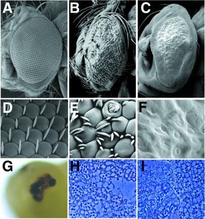
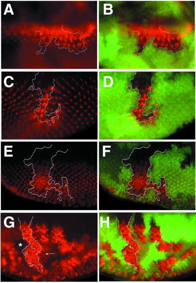
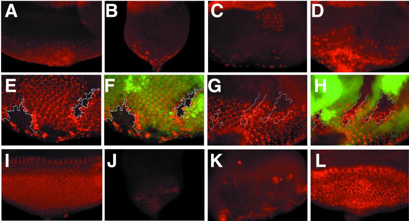
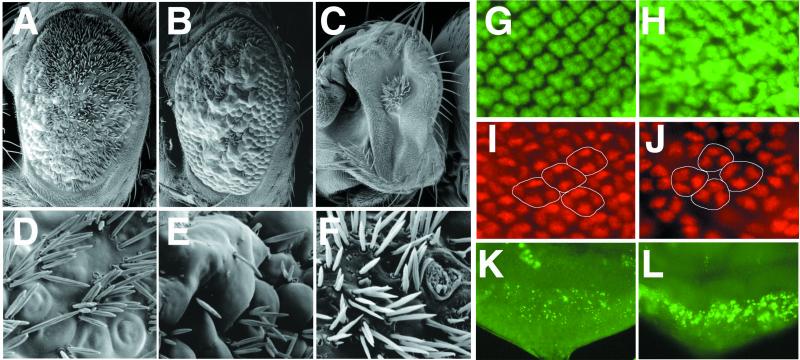
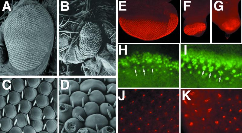
Neuralized (neur) is a neurogenic mutant of Drosophila in which many signaling events mediated by the Notch (N) receptor are disrupted. Here, we analyze the role of neur during eye development. Neur is required in a cell-autonomous fashion to restrict R8 and other photoreceptor fates and is involved in lateral inhibition of interommatidial bristles but is not required for induction of the cone cell fate. The latter contrasts with the absolute requirement for Suppressor of Hairless and the Enhancer of split-Complex for cone cell induction. Using gain-of-function experiments, we further demonstrate that ectopic wild-type and truncated Neur proteins can interfere with multiple N-controlled aspects of eye development, including both neur-dependent and neur-independent processes.


Figures

Figure 1
Figure 1
Phenotypes of neur mutant eyes. (A–F) Scanning electron micrographs (SEM) of adult eyes; (A–C) ≈×150; (D–F) ≈×5,000. (A, D) Wild-type eyes. (B, E) neurA101 mutant eyes generated with ey-FLP. Note tufting of interommatidial bristles, irregular sizes of ommatidia, pitting, and scarring of ommatidia. A strong degree of head macrochaetae tufting around the perimeter of the eye is also apparent. (C, F) neurIF65 mutant eyes generated with ey-FLP. Eyes are bald and smooth; ommatidia lack definition and lenses. Note that head macrochaetae are also absent. (G) Pupal head from an animal aged 45 h after puparium formation containing neurIF65 mutant eyes; a large necrotic patch at the position of the developing eye is present. (H, I) Tangential plastic sections through a neurA101 mutant eye generated by using ey-FLP (H) and an eye containing a neurIF65 mutant clone generated by using hs-FLP (I, mutant clone is left of the dotted line). neurA101 ommatidia frequently have too many rhadomeres, whereas neurIF65 clones have extremely disrupted rhabdomere morphology and numbers; compare with the normal stereotyped arrangement of rhabdomeres in wild-type ommatidia (I, right of the dotted line).
Figure 2
Figure 2
neur is required for lateral inhibition of photoreceptors. Clones of neurIF65 (AF) and neurA101 (G, H) were generated with hs-FLP and are marked by the absence of nuclearly localized GFP detected in green; selected clone boundaries are outlined in white. The expression of different antigens detected in red (A, C, E, G) are shown merged with the GFP clonal marker (B, D, F, H). (A, B) Ato expression normally resolves to single presumptive R8 cells but remains expressed in large clusters within the clone and persists longer than in neighboring wild-type cells. (C, D) Boss is normally expressed in single differentiated R8 cells, but large clusters of Boss-positive cells are found within the clone. Different focal planes are shown in C and D because Boss is apically localized and the clone marker is nuclearly localized; this leads to a slight displacement in the positions of the mutant clone and the phenotypically mutant cells. (E, F) Elav is present in all photoreceptors. A large excess in Elav-positive cells is present within the clone. (G, H) Expression of β-galactosidase in neurA101 homozygous clones. A cell-autonomous increase in the number of β-galactosidase-positive cells is observed within mutant clones; compare with neighboring heterozygous tissue (G, arrow). Note that mutant cells are also homozygous for the enhancer trap and thus produce more β-galactosidase per cell than heterozygous tissue or twin-spot clones, which produce none (G, star).
Figure 3
Figure 3
Requirements of N-pathway members for cone cell induction and E(spl)bHLH expression. Eye discs from wild type (A, I); Su(H)Δ47, P{B} (B, J); eyFLP; E(spl)b32.2, P{gro+} (C, K); eyFLP; neurIF65 (D, L) larvae; eye discs containing hs-FLP induced clones of E(spl)b32.2, P{gro+} (E, F) and neurIF65 (G, H). Note that Su(H) discs are much smaller than eye discs lacking E(spl)-C or neur function. (AD) Expression of Cut, which is found in cone cells as well as adepithelial cells on the surface of the disk; the latter can be distinguished by their larger size and their position in a different focal plane. (B) Su(H) and (C) E(spl)-C mutant discs do not express Cut in cone cells, although staining of adepithelial cells remains; (D) neur mutant discs have an altered pattern of cone cells as marked by Cut. (EH) Staining for Pros, which is present in cone cells and R7. Mutant clones are marked by the absence of GFP and merged images are shown in F and H. (E, F) Clones of the E(spl)-C fail to express Pros in a cell-autonomous fashion, whereas excess Pros staining is observed in wild-type tissue at clone boundaries. (G, H) Clones of neurIF65 express Pros; abnormal pattern of Pros is also observed in wild-type tissue at clone boundaries. (IL) Expression of Mab323, which recognizes 4 of the 7 E(spl)bHLH proteins. Expression is virtually absent in a Su(H) disc (J) and largely eliminated in the E(spl) mutant disc (K); the latter indicates that eyFLP-mediated recombination resulted in an eye disk that is >95% mutant. (L) Expression of Mab323 is maintained in neur mutant disc.
Figure 4
Figure 4
Effect of misexpression of Neur and truncated Neur proteins on adult eye morphology and third instar eye disc development. (AF) SEM of adult eyes of the following genotypes: (A, D) GMR-Gal4; 3xUAS-neur; (B, E) GMR-Gal4; 3xUAS-neurΔRF; (C, F) GMR-Gal4; 2xUAS-neur RING. Misexpression of Neur leads to ommatidial fusions and extreme interommatidial bristle tufting (A, D); compare with wild-type eyes (Fig. 1 A and D). Misexpression of NeurΔRF leads to more pronounced ommatidial fusions and regions of bristle tufting as well as bristle loss (B, E). Misexpression of Neur RING results in a very small disorganized eye (C, F). (GL) Third instar eye discs of the following genotypes: (G, I, K) wild type, (H, J) GMR-Gal4; 3xUAS-neur, (L) GMR-Gal4; 3xUAS-neur RING. (GJ) Posterior regions of the eye disc are shown at a higher magnification than in K and L. Misexpression of Neur causes a mild increase in photoreceptor numbers as marked by Elav (G, H) and decreases the number of cone cells in each ommatidia (circled) as marked by Cut from four to three (I, J). Misexpression of Neur RING results in a strong increase in cell death as marked by acridine orange staining fragments (K, L).
Figure 5
Figure 5
Misexpression of NeurΔRF anterior to the furrow inhibits eye disc growth and lateral inhibition. (AD) SEM of ey-Gal4; 2xUAS-neur (A, C) and ey-Gal4; 2xUAS-neurΔRF; (B, D) adult eyes. (EK) Expression of cell-type specific markers in ey-Gal4/+ (E, H, J), ey-Gal4; 2xUAS-neurΔRF (F, I, K), or Su(H) (G) discs. (EG) Expression of Elav; misexpression of NeurΔRF results in an extremely small retinal field that displays neural hypertrophy (F), similar to the phenotype of Su(H) (G). (H, I) Expression of Ato; resolution of Ato to single R8 cells is incomplete after misexpression of NeurΔRF. A row that has resolved to single Ato-expressing cells in wild-type (H, arrows) maintains Ato in clusters of ≈3 cells (I, arrows) following misexpression of NeurΔRF. (J, K) Expression of Boss; multiple Boss-positive cells are observed in many ommatidia following misexpression of NeurΔRF.

Similar articles

Cited by

References

    1. Brennan C A, Moses K. Cell Mol Life Sci. 2000;57:195–214. - PMC - PubMed
    1. Cagan R L, Ready D F. Genes Dev. 1989;3:1099–1112. - PubMed
    1. Go M J, Eastman D S, Artavanis-Tsakonas S. Development (Cambridge, UK) 1998;125:2031–2040. - PubMed
    1. Papayannopoulos V, Tomlinson A, Panin V M, Rauskolb C, Irvine K D. Science. 1998;281:2031–2034. - PubMed
    1. Dominguez M, de Celis J F. Nature (London) 1998;396:276–278. - PubMed

Publication types