. 2001 May 8;98(10):5776-9.
doi: 10.1073/pnas.101001498.

Involvement of a human gene related to the Drosophila spen gene in the recurrent t(1;22) translocation of acute megakaryocytic leukemia

Affiliations

Involvement of a human gene related to the Drosophila spen gene in the recurrent t(1;22) translocation of acute megakaryocytic leukemia

T Mercher et al. Proc Natl Acad Sci U S A. .

Abstract

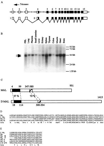
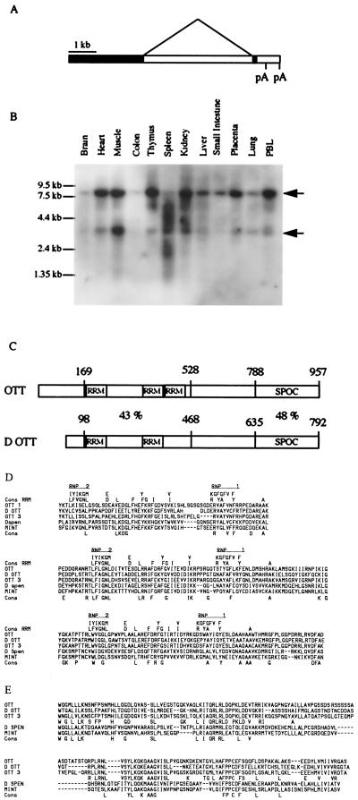
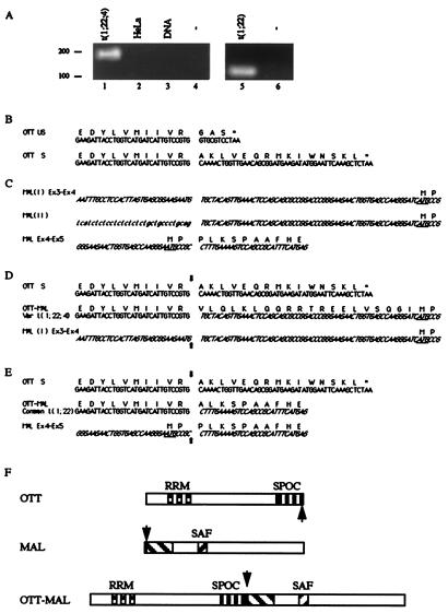
The recurrent t(1;22)(p13;q13) translocation is exclusively associated with infant acute megakaryoblastic leukemia. We have identified the two genes involved in this translocation. Both genes possess related sequences in the Drosophila genome. The chromosome 22 gene (megakaryocytic acute leukemia, MAL) product is predicted to be involved in chromatin organization, and the chromosome 1 gene (one twenty-two, OTT) product is related to the Drosophila split-end (spen) family of proteins. Drosophila genetic experiments identified spen as involved in connecting the Raf and Hox pathways. Because almost all of the sequences and all of the identified domains of both OTT and MAL proteins are included in the predicted fusion protein, the OTT-MAL fusion could aberrantly modulate chromatin organization, Hox differentiation pathways, or extracellular signaling.


Figures

Figure 1
Figure 1
The MAL gene on chromosome 22. (A) The MAL gene comprises 15 exons spanning 226 kb. Predicted coding sequences appear as black boxes and noncoding sequences as empty boxes. (B) Two putative promoters would direct transcription of 4.5 (I) and 4 kb (II) RNA species (shown by arrows) with a variable ratio, depending on the tissues. Translational initiation would occur at the most 5′ in frame ATG located within exon 4. See also Fig. 3C for nucleotide sequences. (C) Representation of the predicted MAL protein along with its closest related Drosophila protein (D mal). The percentage of amino acid identity is shown. The two highest regions of similarity between those two proteins are aligned together with related human predicted proteins. (D) The 95-aa region of unknown function of MAL is aligned with D mal and with human MAL 16 and MAL 17. Amino acids present in at least three of the sequences appear in lowercase letters in the consensus, and those present in the four sequences appear in uppercase letters. The MAL 16 sequences are derived from genomic, EST, and specific RT-PCR products, and MAL 17 sequences are derived from human genomic sequences. (E) The predicted SAF box of MAL is compared with those of D mal, MAL 16, MAL 17. SAF A, SAF B, E1B-AP5, and the SAF consensus sequences are from ref. .
Figure 2
Figure 2
The OTT gene on chromosome 1. (A) Genomic organization of OTT. (B) Northern blot analysis of OTT expression in human tissues by using a probe corresponding to coding sequences. Note that a probe corresponding to the noncoding sequences reacts only with the larger OTT RNA species (data not shown). See also Fig. 3B for nucleotide sequences. (C) Schematic comparison of OTT with its putative Drosophila homolog (D ott). The percentage of amino acid identity is shown. (D) Amino acid alignment of OTT with RNA recognition motif (RRM) consensus (20) and with the similar region of human (OTT3 and MINT) and Drosophila (spen and D ott) proteins. Note that the spacer between RNP2 and RNP1 in the last predicted RRM motif is shorter than the consensus. (E) Amino acid alignment of the same proteins as in D but within the SPOC region. An OTT family consensus is shown in addition to a global one.
Figure 3
Figure 3
OTT-MAL fusion transcripts in infant AML-M7. (A) RT-PCR amplification of OTT-MAL fusion transcripts. A specific OTT-MAL fusion cDNA could be amplified from variant translocation (patient 1, lane 1, 179 bp) or common translocation (patient 2, lane 5, 113 bp) t(1;22), but not from negative control cDNA (HeLa; lane 2), genomic DNA (lane 3), or in the absence of template (lanes 4 and 6). (B) Nucleotide and deduced amino acid sequences of unspliced (US) and spliced (S) normal OTT transcripts. Both species are identifiable in EST and RT-PCR experiments. (C) Nucleotide and deduced amino acid sequences of types I and II normal MAL transcripts. Here and below, chromosome 22 sequences appear in italics, and 5′ untranslated (UT) sequences of type II MAL transcripts absent from type I appear in lowercase letters. (D) Comparison of normal and fused OTT and MAL transcripts, as observed in variant t(1;22;4). The fusion sites are indicated by arrows. The ATG codon underlined in MAL exon 4 sequences is the predicted translational start site of MAL proteins. Stop codons are shown by asterisks. Because the OTT-MAL fusion is in frame and devoid of stop codon, the 5′ UT sequence of MAL exon 4 should be translated in this case. (E) Comparison of normal and fused OTT and MAL transcripts, as observed in common t(1;22). (F) Representation of predicted normal and fusion proteins, as deduced from the common t(1;22) structural analysis. Interspecies conserved regions are indicated, and fusion points are shown by arrows.

References

    1. Rabbitts T H. Nature (London) 1994;372:143–149. - PubMed
    1. Rowley J D. Annu Rev Genet. 1998;32:495–519. - PubMed
    1. Look A T. Science. 1997;278:1059–1064. - PubMed
    1. Mann R S, Chan S K. Trends Genet. 1996;12:258–262. - PubMed
    1. Rowley J D. Semin Cancer Biol. 1993;4:377–385. - PubMed

Publication types