

Sensorimotor mapping of the human cerebellum: fMRI evidence of somatotopic organization
- PMID: 11346886
- PMCID: PMC6871814
- DOI: 10.1002/hbm.1025
Sensorimotor mapping of the human cerebellum: fMRI evidence of somatotopic organization
Abstract
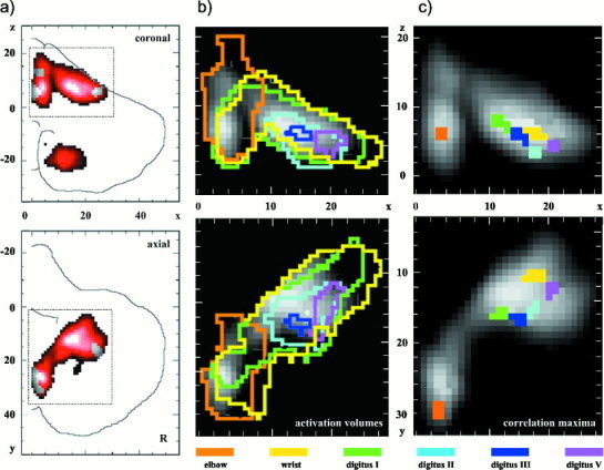
Functional magnetic resonance imaging (fMRI) was employed to determine areas of activation in the cerebellar cortex in 46 human subjects during a series of motor tasks. To reduce the variance due to differences in individual anatomy, a specific transformational procedure for the cerebellum was introduced. The activation areas for movements of lips, tongue, hands, and feet were determined and found to be sharply confined to lobules and sublobules and their sagittal zones in the rostral and caudal spino-cerebellar cortex. There was a clear symmetry mirroring at the midline. The activation mapped as two distinct homunculoid representations. One, a more extended representation, was located upside down in the superior cerebellum, and a second one, doubled and smaller, in the inferior cerebellum. The two representations were remarkably similar to those proposed by Snider and Eldred [1951] five decades ago. In the upper representation, an intralimb somatotopy for the right elbow, wrist, and fingers was revealed. The maps seem to confirm earlier electrophysiological findings of sagittal zones in animals. They differ, however, from micromapping reports on fractured somatotopic maps in the cerebellar cortex of mammals. We assume that the representations that we observed are not solely the result of spatial integration of hemodynamic events underlying the fMRI method and may reflect integration of afferent peripheral and central information in the cerebellar cortex.
Copyright 2001 Wiley-Liss, Inc.
Figures


References
-
- Allen G, Buxton RB, Wong EC, Courchesne E (1997): Attentional activation of the cerebellum independent of motor involvement. Science 275: 1940–1942. - PubMed
-
- Bandettini PA, Jesmanowicz A, Wong EC, Hyde JS (1993): Processing strategies for time‐course data sets in functional MRI of the human brain. Magn Reson Med 30: 161–173. - PubMed
-
- Bower JM, Woolston DC (1983): Congruence of spatial organization of tactile projections to granule cell and Purkinje cell layers of the cerebellar hemispheres of the albino rat: vertical organization of cerebellar cortex. J Neurophysiol 49: 745–766. - PubMed
-
- Bower JM (1997): Is the cerebellum sensory for motor's sake, or motor for sensory's sake: the view from the whiskers of a rat? Prog Brain Res 114: 463–96. - PubMed
-
- Braitenberg V, Atwood RP (1958): Morphological observations on the cerebellar cor‐tex. J Comp Neurol 109: 1–27. - PubMed
Publication types
MeSH terms
LinkOut - more resources
Full Text Sources
