

Regulation of OspE-related, OspF-related, and Elp lipoproteins of Borrelia burgdorferi strain 297 by mammalian host-specific signals
- PMID: 11349022
- PMCID: PMC98350
- DOI: 10.1128/IAI.69.6.3618-3627.2001
Regulation of OspE-related, OspF-related, and Elp lipoproteins of Borrelia burgdorferi strain 297 by mammalian host-specific signals
Abstract
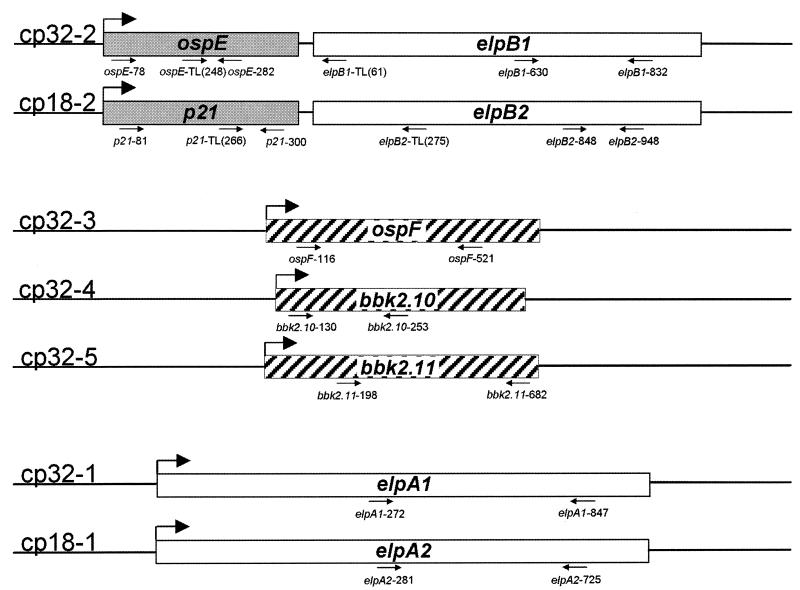
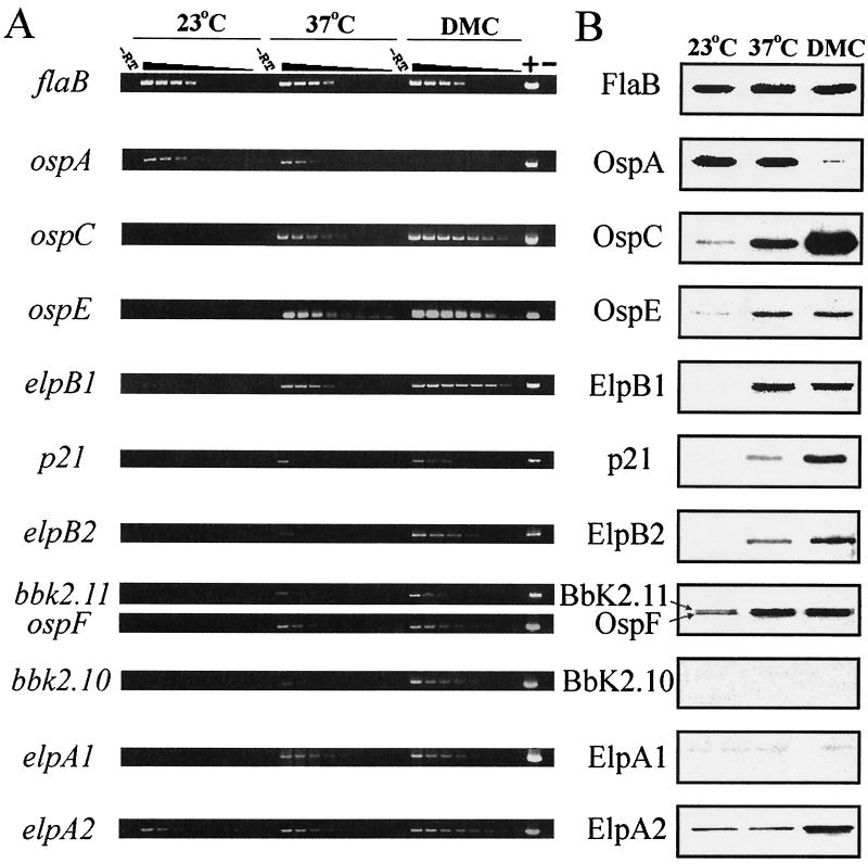
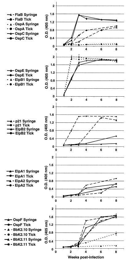
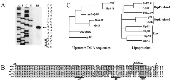
In previous studies we have characterized the cp32/18 loci in Borrelia burgdorferi 297 which encode OspE and OspF orthologs and a third group of lipoproteins which possess OspE/F-like leader peptides (Elps). To further these studies, we have comprehensively analyzed their patterns of expression throughout the borrelial enzootic cycle. Serial dilution reverse transcription-PCR analysis indicated that although a shift in temperature from 23 to 37 degrees C induced transcription for all nine genes analyzed, this effect was often markedly enhanced in mammalian host-adapted organisms cultivated within dialysis membrane chambers (DMCs) implanted within the peritoneal cavities of rats. Indirect immunofluorescence assays performed on temperature-shifted, in vitro-cultivated spirochetes and organisms in the midguts of unfed and fed ticks revealed distinct expression profiles for many of the OspE-related, OspF-related, and Elp proteins. Other than BbK2.10 and ElpA1, all were expressed by temperature-shifted organisms, while only OspE, ElpB1, OspF, and BbK2.11 were expressed in the midguts of fed ticks. Additionally, although mRNA was detected for all nine lipoprotein-encoding genes, two of these proteins (BbK2.10 and ElpA1) were not expressed by spirochetes cultivated in vitro, within DMCs, or by spirochetes within tick midguts. However, the observation that B. burgdorferi-infected mice generated specific antibodies against BbK2.10 and ElpA1 indicated that these antigens are expressed only in the mammalian host and that a form of posttranscriptional regulation is involved. Analysis of the upstream regions of these genes revealed several differences between their promoter regions, the majority of which were found in the -10 and -35 hexamers and the spacer regions between them. Also, rather than undergoing simultaneous upregulation during tick feeding, these genes and the corresponding lipoproteins appear to be subject to progressive recruitment or enhancement of expression as B. burgdorferi is transmitted from its tick vector to the mammalian host. These findings underscore the potential relevance of these molecules to the pathogenic events of early Lyme disease.
Figures
Similar articles
-
Changes in temporal and spatial patterns of outer surface lipoprotein expression generate population heterogeneity and antigenic diversity in the Lyme disease spirochete, Borrelia burgdorferi.Infect Immun. 2002 Jul;70(7):3468-78. doi: 10.1128/IAI.70.7.3468-3478.2002. Infect Immun. 2002. PMID: 12065486 Free PMC article.
-
Molecular and evolutionary analysis of Borrelia burgdorferi 297 circular plasmid-encoded lipoproteins with OspE- and OspF-like leader peptides.Infect Immun. 1999 Mar;67(3):1526-32. doi: 10.1128/IAI.67.3.1526-1532.1999. Infect Immun. 1999. PMID: 10024606 Free PMC article.
-
Temporal pattern of Borrelia burgdorferi p21 expression in ticks and the mammalian host.J Clin Invest. 1997 Mar 1;99(5):987-95. doi: 10.1172/JCI119264. J Clin Invest. 1997. PMID: 9062357 Free PMC article.
-
New Insights Into CRASP-Mediated Complement Evasion in the Lyme Disease Enzootic Cycle.Front Cell Infect Microbiol. 2020 Jan 30;10:1. doi: 10.3389/fcimb.2020.00001. eCollection 2020. Front Cell Infect Microbiol. 2020. PMID: 32083019 Free PMC article. Review.
-
Lyme borreliosis spirochete Erp proteins, their known host ligands, and potential roles in mammalian infection.Int J Med Microbiol. 2008 Sep 1;298 Suppl 1(Suppl 1):257-67. doi: 10.1016/j.ijmm.2007.09.004. Epub 2008 Jan 11. Int J Med Microbiol. 2008. PMID: 18248770 Free PMC article. Review.
Cited by
-
Host cell heparan sulfate glycosaminoglycans are ligands for OspF-related proteins of the Lyme disease spirochete.Cell Microbiol. 2015 Oct;17(10):1464-76. doi: 10.1111/cmi.12448. Epub 2015 May 13. Cell Microbiol. 2015. PMID: 25864455 Free PMC article.
-
BpaB and EbfC DNA-binding proteins regulate production of the Lyme disease spirochete's infection-associated Erp surface proteins.J Bacteriol. 2012 Feb;194(4):778-86. doi: 10.1128/JB.06394-11. Epub 2011 Dec 9. J Bacteriol. 2012. PMID: 22155777 Free PMC article.
-
Autophagy modulates Borrelia burgdorferi-induced production of interleukin-1β (IL-1β).J Biol Chem. 2013 Mar 22;288(12):8658-8666. doi: 10.1074/jbc.M112.412841. Epub 2013 Feb 5. J Biol Chem. 2013. PMID: 23386602 Free PMC article.
-
Demonstration of the genetic stability and temporal expression of select members of the lyme disease spirochete OspF protein family during infection in mice.Infect Immun. 2001 Aug;69(8):4831-8. doi: 10.1128/IAI.69.8.4831-4838.2001. Infect Immun. 2001. PMID: 11447157 Free PMC article.
-
Borrelia burgdorferi erp (ospE-related) gene sequences remain stable during mammalian infection.Infect Immun. 2002 Sep;70(9):5307-11. doi: 10.1128/IAI.70.9.5307-5311.2002. Infect Immun. 2002. PMID: 12183589 Free PMC article.
References
-
- Akada J K, Shirai M, Takeuchi H, Tsuda M, Nakazawa T. Identification of the urease operon in Helicobacter pylori and its control by mRNA decay in response to pH. Mol Microbiol. 2000;36:1071–1084. - PubMed
-
- Akins D R, Porcella S F, Popova T G, Shevchenko D, Baker S I, Li M, Norgard M V, Radolf J D. Evidence for in vivo but not in vitro expression of a Borrelia burgdorferi outer surface protein F (OspF) homolog. Mol Microbiol. 1995;18:507–520. - PubMed
-
- Anderson D M, Schneewind O. A mRNA signal for the type III secretion of Yop proteins by Yersinia enterocolitica. Science. 1997;278:1140–1143. - PubMed
Publication types
MeSH terms
Substances
Grants and funding
LinkOut - more resources
Full Text Sources
Medical
