. 2001 Jun;69(6):3995-4006.
doi: 10.1128/IAI.69.6.3995-4006.2001.

Intracellular survival of Brucella spp. in human monocytes involves conventional uptake but special phagosomes

Affiliations

Intracellular survival of Brucella spp. in human monocytes involves conventional uptake but special phagosomes

M G Rittig et al. Infect Immun. 2001 Jun.

Abstract

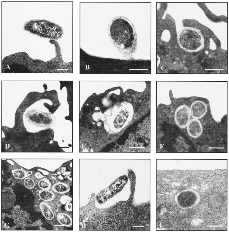
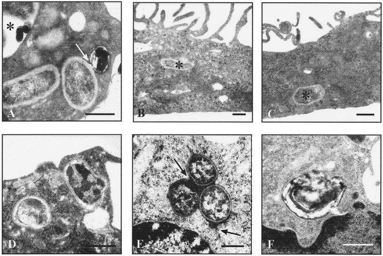
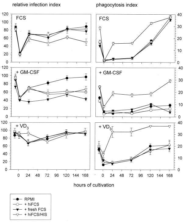
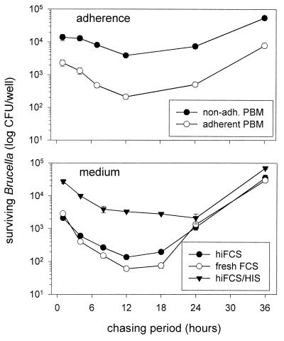
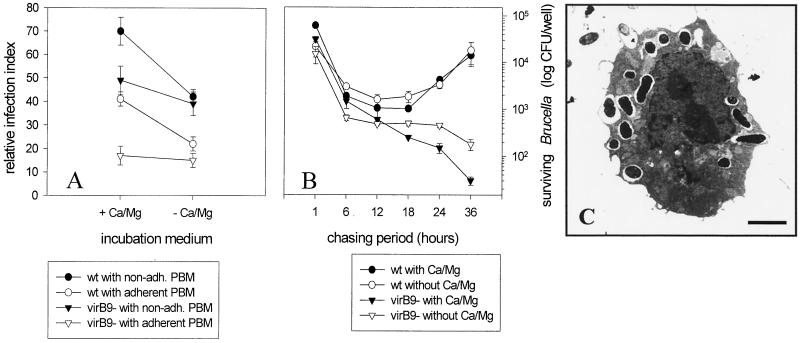
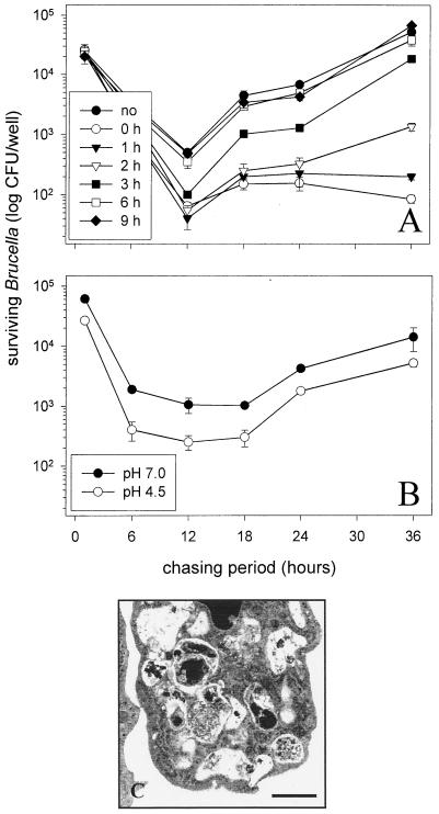
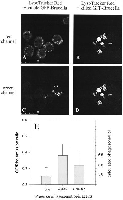
Brucella spp. are facultative intracellular parasites of various mammals, including humans, typically infecting lymphoid as well as reproductive organs. We have investigated how B. suis and B. melitensis enter human monocytes and in which compartment they survive. Peripheral blood monocytes readily internalized nonopsonized brucellae and killed most of them within 12 to 18 h. The presence of Brucella-specific antibodies (but not complement) increased the uptake of bacteria without increasing their intracellular survival, whereas adherence of the monocytes or incubation in Ca(2+)- and Mg(2+)-free medium reduced the uptake. Engulfment of all Brucella organisms (regardless of bacterial viability or virulence) initially resulted in phagosomes with tightly apposed walls (TP). Most TP were fully fusiogenic and matured to spacious phagolysosomes containing degraded bacteria, whereas some TP (more in monocyte-derived macrophages, HeLa cells, and CHO cells than in monocytes) remained tightly apposed to intact bacteria. Immediate treatment of infected host cells with the lysosomotropic base ammonium chloride caused a swelling of all phagosomes and a rise in the intraphagosomal pH, abolishing the intracellular survival of Brucella. These results indicate that (i) human monocytes readily internalize Brucella in a conventional way using various phagocytosis-promoting receptors, (ii) the maturation of some Brucella phagosomes is passively arrested between the steps of acidification and phagosome-lysosome fusion, (iii) brucellae are killed in maturing but not in arrested phagosomes, and (iv) survival of internalized Brucella depends on an acidic intraphagosomal pH and/or close contact with the phagosomal wall.


Figures

FIG. 1
FIG. 1
Ultrastructural observations on the phagocytosis of Brucella by human monocytes and epithelioid cells. Freshly isolated monocytes (A to G) or epithelioid cells (H and I) were challenged with nonopsonized B. suis 1330 for 15 min and chased for up to 24 h. Bars, 0.2 μm. Brucellae attached to various parts of the plasma membrane, such as cell surface projections (A) or the plain cell surface (B). The emerging phagocytic cups which formed within the first minutes had either continuous (C) or focal (D) contact with the bacteria. The resulting phagosomes either were spacious (E) or had tightly apposed walls (F). Both types of phagosomes showed fusion with small intracellular vesicles, with the fusion events for the latter type being more numerous after longer chasing periods (G) (asterisks indicate phagosome-lysosome fusion; chase was for 2 h). Uptake of Brucella by HeLa or CHO cells also took place via the usual phagocytic mechanisms (H) (HeLa cells) and resulted in membrane-bound compartments which predominantly had tightly apposed walls (I) (CHO cells).
FIG. 2
FIG. 2
Ultrastructural observations on Brucella-bearing phagosomes in human monocytes. Monocyte-derived macrophages (A to C) or freshly isolated monocytes (D to F) were challenged with B. suis 1330 for 15 min and chased for up to 8 h. Bars, 0.2 μm. In MDM differentiated for 7 days in the presence of FCS without additions (A) or with either 100 nM VD3 (B) or 500 U of GM-CSF per ml (C), brucellae were located almost exclusively in TP (asterisks in panels B and C). Especially after longer chasing periods, Brucella-bearing phagosomes occasionally contained electron-dense membrane remnants, as indicated for an SP (asterisk in panel A) and for a vesicle fusing with a TP (arrow in panel A). Formation of TP did not depend on bacterial viability or virulence, as shown with two TP enclosing heat-killed brucellae (D). Preloading endosomal compartments of the host cells with electron-dense markers revealed that TP were able to fuse with early but not late endosomes (E), with three TP grouped around a centrally placed early endosome (upper arrow) and one TP fusing with an additional early endosome (lower arrow). Osmiophilic precipitations of polymerized DAB, indicative of oxidative burst, were occasionally present in some SP but not in TP (F), even when using antibody-opsonized brucellae.
FIG. 3
FIG. 3
Uptake of differently opsonized brucellae by human monocytes and MDM. Uptake of Brucella was compared for freshly isolated, nonadherent monocytes (pre-0-h time point) and monocytes which were allowed to adhere for 1 h (0-h time point) and kept in culture for up to 7 days (24-, 72-, 120-, and 168-h time points). At each time point indicated, phagocyte aliquots were challenged with GFP-expressing B. suis for 20 min and chased for another 30 min. The effects of different supplements to the monocyte culture medium (10% hiFCS [FCS]), FCS with 500 U of GM-CSF per ml [+ GM-CSF]), or FCS with 100 nM VD3 [+ VD3]) and different challenging media (RPMI alone [RPMI] or RPMI containing either 10% hiFCS [+ hiFCS], fresh FCS [+ fresh FCS], or hiFCS with 5 μl of human anti-Brucella immune serum [+ hiFCS/HIS]) were compared. Infection was quantified by count of monocyte-associated fluorescent bacteria. Results are expressed as means ± standard deviations for duplicate samples from one of two experiments with monocytes from different donors which gave comparable results.
FIG. 4
FIG. 4
Intracellular survival of differently opsonized brucellae following uptake by nonadherent and adherent human monocytes. Freshly isolated PBM were challenged with B. suis for 20 min and chased for 1 to 36 h. Viable bacteria reisolated from osmotically lysed PBM were determined by count of CFU. (Top panel) Survival of nonopsonized brucellae internalized by nonadherent (non-adh.) and adherent PBM. (Bottom panel) Survival of brucellae internalized by nonadherent PBM in the presence of either hiFCS, fresh FCS, or 5 μl of human anti-Brucella immune serum per ml in hiFCS (hiFCS/HIS). Results are expressed as means ± standard deviations for duplicate samples from one of three experiments with monocytes from different donors which gave comparable results.
FIG. 5
FIG. 5
Role of Ca2+, monocyte adherence, and the bacterial virB9 gene cluster in uptake and intracellular survival of Brucella. (A and B) Freshly isolated PBM were challenged with nonopsonized GFP-expressing wild-type (wt) B. suis or a virB9 mutant for 20 min and chased for another 30 min. Results are expressed as means ± standard deviations for duplicate samples from one out of three experiments with monocytes from different donors which gave comparable results. (A) Uptake. Counts of fluorescent bacteria internalized by nonadherent (non-adh.) or adherent PBM in the presence (+ Ca/Mg) or absence (− Ca/Mg) of external Ca2+ and Mg2+ are shown. (B) Survival. Counts of CFU of bacteria reisolated from osmotically lysed PBM following uptake in the presence or absence of external Ca2+ and Mg2+ are shown. (C) Electron micrograph of a PBM treated with 2 μM thapsigargin for 5 min before challenge with wt B. suis (15-min pulse, 60-min chase) in the presence of external Ca2+ and Mg2+. Thapsigargin-induced depletion of the internal Ca2+ stores did not affect the uptake of Brucella but did affect the centripetal transport of the phagosomes, as indicated by the predominantly peripheral localization of the phagosomes. The walls of the phagosomes show no tight apposition. Bar, 7 μm.
FIG. 6
FIG. 6
Effect of ammonium chloride (A) and acidic shock (B) on the intracellular survival of Brucella in human monocytes. Freshly isolated PBM were challenged with nonopsonized B. suis for 20 min and chased for 1 to 36 h. Viable bacteria reisolated from osmotically lysed monocytes were determined by count of CFU. Results are expressed as means ± standard deviations for duplicate samples from one out of three experiments with monocytes from different donors which gave comparable results. (A) Compared is the survival of B. suis in the absence (no) or presence of 30 mM ammonium chloride added either immediately (0 h) at the onset of chase or up to 9 h later. (B) Compared is the survival of B. suis kept at either neutral or acidic pH for 4 h prior to infection. (C) Electron micrograph of a Brucella-infected monocyte chased for 8 h in the continuous presence of ammonium chloride. All Brucella-bearing phagosomes are spacious, and the internalized bacteria are degraded. Bar, 1 μm.
FIG. 7
FIG. 7
Measurement of phagosomal pH in Brucella-infected murine macrophage-like cells. (A to D) J774.A1 cells were challenged with antibody-opsonized, viable (A and C) or heat-killed (B and D) GFP-expressing Brucella for 45 min and chased for another 90 min in the presence of the acidophilic reagent LysoTracker Red. Depicted are paired images in the red (A and B) and green (C and D) channels for visualization of LysoTracker Red and GFP-expressing Brucella, respectively. Even at the high concentration of 1 μM used here, LysoTracker Red colocalizes only weakly and inconsistently with viable Brucella but to the full extent with heat-killed Brucella. These features were observed throughout all infected cells in duplicate experiments. (E) J774.A1 cells were challenged with antibody-opsonized, viable CF- and Rho-labeled Brucella for 45 min and chased for another 90 min in the absence or presence of either 100 nM bafilomycin A1 (BAF) or 30 mM ammonium chloride. Results are expressed as means ± standard deviations for four pairs of CF-Rho images of duplicate experiments; the phagosomal pH was calculated from an in situ calibration curve of the CF/Rho emission ratio versus buffers with defined pHs. Heat-killed brucellae gave the same results as viable ones (data not shown).

Similar articles

Cited by

References

    1. Ackermann M, Cheville N, Deyoe B. Bovine ileal dome lymphoepithelial cells: endocytosis and transport of Brucella abortus strain 19. Vet Pathol. 1988;25:28–35. - PubMed
    1. Anderson T, Cheville N. Ultrastructural morphometric analysis of Brucella abortus-infected trophoblasts in experimental placentitis. Bacterial replication occurs in rough endoplasmic reticulum. Am J Pathol. 1986;124:226–237. - PMC - PubMed
    1. Arenas G N, Staskevich A S, Aballay A, Mayorga L S. Intracellular trafficking of Brucella abortus in J774 macrophages. Infect Immun. 2000;68:4255–4263. - PMC - PubMed
    1. Barclay N, Brown M, Law S, McKnight J, Tomlinson M, van der Merwe P. The leucocyte antigen facts book. 2nd ed. San Diego, Calif: Academic Press; 1997.
    1. Bounous D, Enright F, Gossett K, Berry C. Phagocytosis, killing, and oxidant production by bovine monocyte-derived macrophages upon exposure to Brucella abortus strain 2308. Vet Immunol Immunopathol. 1993;37:243–256. - PubMed

Publication types

MeSH terms

LinkOut - more resources