. 2001 Jun;69(6):4027-33.
doi: 10.1128/IAI.69.6.4027-4033.2001.

EspG, a novel type III system-secreted protein from enteropathogenic Escherichia coli with similarities to VirA of Shigella flexneri

Affiliations

EspG, a novel type III system-secreted protein from enteropathogenic Escherichia coli with similarities to VirA of Shigella flexneri

S J Elliott et al. Infect Immun. 2001 Jun.

Abstract

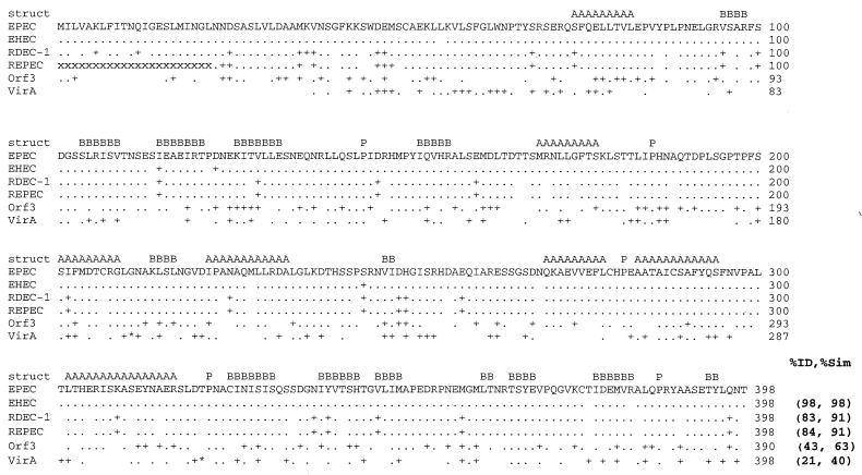
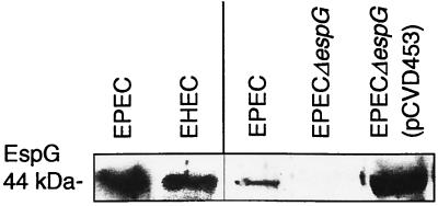
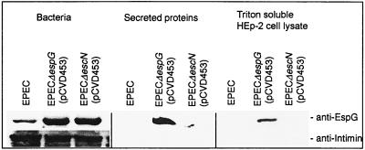
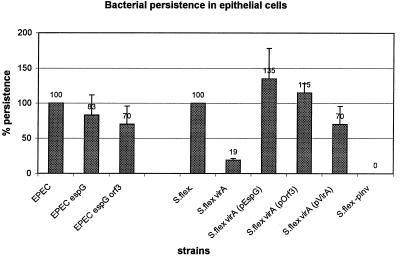
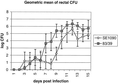
The function of the rorf2 gene located on the locus of enterocyte effacement (LEE) pathogenicity island of enteropathogenic Escherichia coli (EPEC) has not been described. We report that rorf2 encodes a novel protein, named EspG, which is secreted by the type III secretory system and which is translocated into host epithelial cells. EspG is homologous with Shigella flexneri protein VirA, and the cloned espG (rorf2) gene can rescue invasion in a Shigella virA mutant, indicating that these proteins are functionally equivalent in Shigella. An EPEC espG mutant had no apparent defects in in vitro assays of virulence phenotypes, but a rabbit diarrheagenic E. coli strain carrying a mutant espG showed diminished intestinal colonization and yet diarrheal attack rates similar to those of the wild type. A second EspG homolog, Orf3, is encoded on the EspC pathogenicity islet. The cloned orf3 gene could also rescue invasion in a Shigella virA mutant, but an EPEC espG orf3 double mutant was not diminished in any tested in vitro assays for EPEC virulence factors. Our results indicate that EspG plays an accessory but as yet undefined role in EPEC virulence that may involve intestinal colonization.


Figures

FIG. 1
FIG. 1
Alignment of EspG from EPEC O127: H7 (EPEC) with EspG from EHEC O157: H7 (EHEC), rabbit pathogen RDEC-1, REPEC strain 83/39 (REPEC), Orf3 from the EspC pathogenicity islet, and VirA from S. flexneri. Numbers at the end of the line, amino acid numbers; period, identical amino acids; +, similar but nonidentical amino acids; blank space, nonhomologous amino acids. The DNA sequence encoding the first 22 amino acids (aa) of REPEC VirA is not known, and each of these missing amino acids is indicated with an “x.” Asterisks (within the VirA sequence), areas where VirA contains extra amino acids not found within EspG, including an insertion of 5 aa into the region corresponding to aa 210 and 211 in EspG and a 15-aa insertion between aa 320 and 321 in EspG. The predicted consensus secondary structures (struct) conserved between EspG and VirA are denoted A (α-helix), B (β sheet), and P (proline; indicating a turn). The percent identity and percent similarity (%ID and %Sim, respectively) to the sequence of EPEC EspG are listed at the end of the alignment.
FIG. 2
FIG. 2
Western blot with anti-EspG antiserum on whole-cell lysates. Exposure times: lanes 1 and 2 (numbering from the left), 3 min; lanes 3 to 5, 15 s. Antiserum recognized the 44-kDa EspG protein in whole-cell lysates of wild-type EPEC E2348/69 (lanes 1 and 3), EHEC (lane 2), and EPEC E2348/69 espG (pCVD453) (lane 5) but not in that of EPEC E2348/69 espG (lane 4). Comparison of relative amounts of EspG produced by EPEC, the espG mutant, and the complemented mutant (lanes 3 to 5) demonstrates that EspG is produced in small amounts by wild-type EPEC and that the complement overproduces EspG.
FIG. 3
FIG. 3
Type III secretion of EspG into supernatants and translocation into HEp-2 cells. Western blots with EspG antiserum indicate that EspG is produced by EPEC E2348/69 espG (pCVD453), EPEC E2348/69 escN (pCVD453), and in lesser amounts by wild-type EPEC E2348/69. EspG is observed in the supernatant and in the Triton X-100-soluble fraction of HEp-2 cells infected with EPEC E2348/69 espG (pCVD453) but not EPEC E2348/69 escN (pCVD453), indicating that secretion and translocation are dependent on type III secretion. Intimin is observed in whole cells but not in the Triton X-100-soluble fraction, indicating that the Triton X-100-soluble fraction is not contaminated with bacteria, and so EspG present in that fraction must be due to translocation and not contamination.
FIG. 4
FIG. 4
Intracellular persistence of bacteria within HeLa cells as a measure of intracellular survival and/or replication, according to the protocol of Anderson et al. (2), and expressed as percentages of that for wild-type EPEC or S. flexneri (S.flex). The plasmid-free variant of S. flexneri (S.flex-pInv) was used as a negative control. Relative scores are above the bars.
FIG. 5
FIG. 5
Geometric mean of the CFU count from rectal swabs, as a measure of fecal CFU, from rabbits orally inoculated with wild-type REPEC strain 83/39 or espG mutant SE1090. Error bars, standard errors.

Similar articles

Cited by

References

    1. Adams L M, Simmons C P, Rezmann L, Strugnell R A, Robins-Browne R M. Identification and characterization of a K88- and CS31A-like operon of a rabbit enteropathogenic Escherichia coli strain which encodes fimbriae involved in the colonization of rabbit intestine. Infect Immun. 1997;65:5222–5230. - PMC - PubMed
    1. Anderson R J, Pasetti M F, Sztein M B, Levine M M, Noriega F R. ΔguaBA attenuated Shigella flexneri 2a strain CVD 1204 as a Shigella vaccine and as a live mucosal delivery system for fragment C of tetanus toxin. Vaccine. 2000;18:2193–2202. - PubMed
    1. Deibel C, Krämer S, Chakraborty T, Ebel F. EspE, a novel secreted protein of attaching and effacing bacteria, is directly translocated into infected host cells where it appears as a tyrosine-phosphorylated 90 kDa protein. Mol Microbiol. 1998;28:463–474. - PubMed
    1. Demers B, Sansonetti P J, Parsot C. Induction of type III secretion in Shigella flexneri is associated with differential control of transcription of genes encoding secreted proteins. EMBO J. 1998;17:2894–2903. - PMC - PubMed
    1. Donnenberg S M, Donohue-Rolfe A, Keusch G T. Epithelial cell invasion: an overlooked property of enteropathogenic Escherichia coli (EPEC) associated with the EPEC adherence factor. J Infect Dis. 1989;160:452–459. - PubMed

Publication types

MeSH terms