Induction of autoimmune valvular heart disease by recombinant streptococcal m protein
- PMID: 11349078
- PMCID: PMC98471
- DOI: 10.1128/IAI.69.6.4072-4078.2001
Induction of autoimmune valvular heart disease by recombinant streptococcal m protein
Abstract
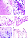
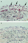
Rheumatic heart disease is an autoimmune sequela of group A streptococcal infection. Previous studies have established that streptococcal M protein is structurally and immunologically similar to cardiac myosin, a well-known mediator of inflammatory heart disease. In this study, we investigated the hypothesis that streptococcal M protein could produce inflammatory valvular heart lesions similar to those seen in rheumatic fever (RF). Fifty percent (3 of 6) of Lewis rats immunized with recombinant type 6 streptococcal M protein (rM6) developed valvulitis as well as focal lesions of myocarditis. Valvular lesions initiated at the valve surface endothelium spread into the valve. Anitschkow cells and verruca-like lesions were present. T cells from rM6-immunized rats proliferated in the presence of purified cardiac myosin, but not skeletal myosin. A T-cell line produced from rM6-treated rats proliferated in the presence of cardiac myosin and rM6 protein. The study demonstrates that the Lewis rat is a model of valvular heart disease and that streptococcal M protein can induce an autoimmune cell-mediated immune attack on the heart valve in an animal model. The data support the hypothesis that a bacterial antigen can break immune tolerance in vivo, an important concept in autoimmunity.
Figures


Similar articles
-
Induction of myocarditis and valvulitis in lewis rats by different epitopes of cardiac myosin and its implications in rheumatic carditis.Am J Pathol. 2002 Jan;160(1):297-306. doi: 10.1016/S0002-9440(10)64373-8. Am J Pathol. 2002. PMID: 11786423 Free PMC article.
-
Induction of autoimmune valvulitis in Lewis rats following immunization with peptides from the conserved region of group A streptococcal M protein.J Autoimmun. 2003 May;20(3):211-7. doi: 10.1016/s0896-8411(03)00026-x. J Autoimmun. 2003. PMID: 12753806
-
T cell mimicry in inflammatory heart disease.Mol Immunol. 2004 Feb;40(14-15):1121-7. doi: 10.1016/j.molimm.2003.11.023. Mol Immunol. 2004. PMID: 15036918 Review.
-
Group A streptococcal M-protein specific antibodies and T-cells drive the pathology observed in the rat autoimmune valvulitis model.Autoimmunity. 2019 Mar;52(2):78-87. doi: 10.1080/08916934.2019.1605356. Epub 2019 May 7. Autoimmunity. 2019. PMID: 31062619
-
Cardiac contractile proteins and autoimmune myocarditis.Mol Cell Biochem. 1993 Feb 17;119(1-2):67-71. doi: 10.1007/BF00926855. Mol Cell Biochem. 1993. PMID: 8455588 Review.
Cited by
-
In Search of the Holy Grail: A Specific Diagnostic Test for Rheumatic Fever.Front Cardiovasc Med. 2021 May 14;8:674805. doi: 10.3389/fcvm.2021.674805. eCollection 2021. Front Cardiovasc Med. 2021. PMID: 34055941 Free PMC article.
-
Triggers of Inflammatory Heart Disease.Front Cell Dev Biol. 2020 Mar 24;8:192. doi: 10.3389/fcell.2020.00192. eCollection 2020. Front Cell Dev Biol. 2020. PMID: 32266270 Free PMC article. Review.
-
Protection against or triggering of Type 1 diabetes? Different roles for viral infections.Expert Rev Clin Immunol. 2011 Jan;7(1):45-53. doi: 10.1586/eci.10.91. Expert Rev Clin Immunol. 2011. PMID: 21162649 Free PMC article. Review.
-
Streptococcus and rheumatic fever.Curr Opin Rheumatol. 2012 Jul;24(4):408-16. doi: 10.1097/BOR.0b013e32835461d3. Curr Opin Rheumatol. 2012. PMID: 22617826 Free PMC article. Review.
-
Genes, autoimmunity and pathogenesis of rheumatic heart disease.Ann Pediatr Cardiol. 2011 Jan;4(1):13-21. doi: 10.4103/0974-2069.79617. Ann Pediatr Cardiol. 2011. PMID: 21677799 Free PMC article.
References
-
- Adderson E E, Shikhman A R, Ward K E, Cunningham M W. Molecular analysis of polyreactive monoclonal antibodies from rheumatic carditis: human anti-N-acetyl-glucosamine/anti-myosin antibody V region genes. J Immunol. 1998;161:2020–2031. - PubMed
-
- Antone S M, Adderson E E, Mertens N M J, Cunningham M W. Molecular analysis of V gene sequences encoding cytotoxic anti-streptococcal/anti-myosin monoclonal antibody 36.2.2 that recognizes the heart cell surface protein laminin. J Immunol. 1997;159:5422–5430. - PubMed
-
- Ayoub E M. Resurgence of rheumatic fever in the United States. Postgrad Med. 1992;92:133–142. - PubMed
-
- Coligan J E, Kruisbeck A D, Margulies D H, Shevach E M, Strober W. Current protocols in immunology. 1, section 3.12. New York, N.Y: Greene Publishing and Wiley-Interscience; 1994.
Publication types
MeSH terms
Substances
Grants and funding
LinkOut - more resources
Full Text Sources
Medical

