. 2001 May 15;20(10):2394-403.
doi: 10.1093/emboj/20.10.2394.

Evolutionarily divergent electron donor proteins interact with P450MT2 through the same helical domain but different contact points

Affiliations

Evolutionarily divergent electron donor proteins interact with P450MT2 through the same helical domain but different contact points

H K Anandatheerthavarada et al. EMBO J. .

Abstract

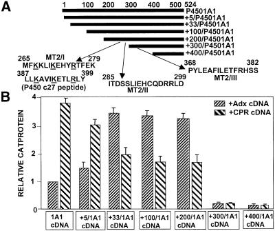
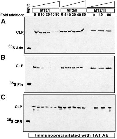
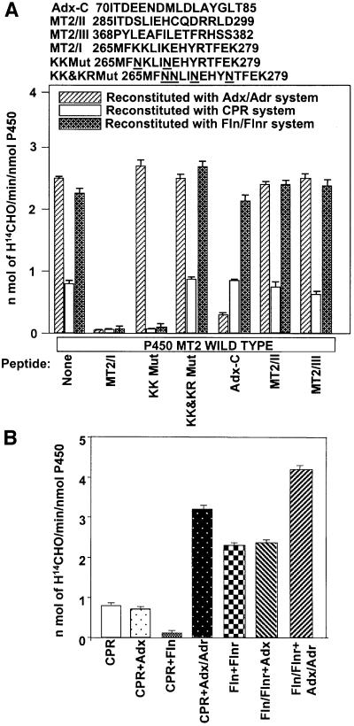
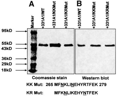
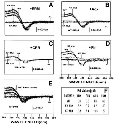
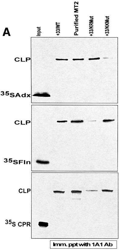
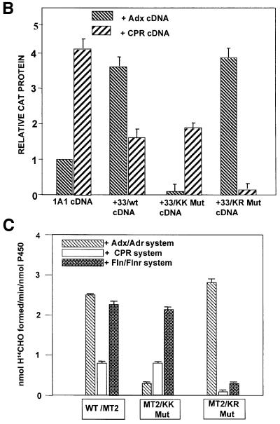
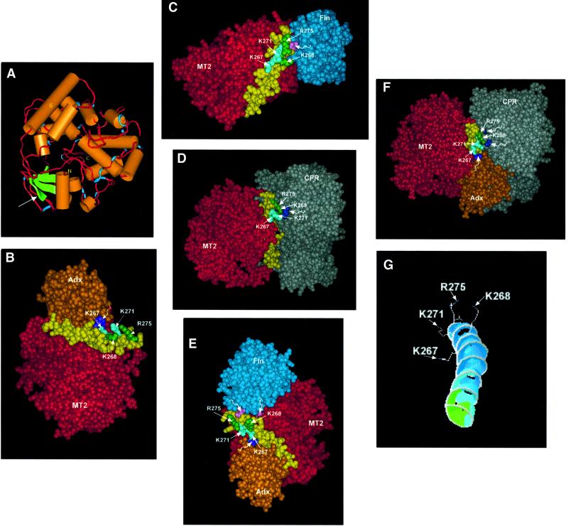
We have investigated the sites of N-terminally truncated cytochrome P4501A1 targeted to mitochondria (P450MT2) which interact with adrenodoxin (Adx), cytochrome P450 reductase (CPR) and bacterial flavodoxin (Fln). The binding site was mapped by a combination of in vitro mutagenesis, in vivo screening with a mammalian two-hybrid system, spectral analysis, reconstitution of enzyme activity and homology-based structural modeling. Our results show that part of an aqueous accessible helix (putative helix G, residues 264-279) interacts with all three electron donor proteins. Mutational studies revealed that Lys267 and Lys271 are crucial for binding to Adx, while Lys268 and Arg275 are important for binding to CPR and FLN: Additive effects of different electron donor proteins on enzyme activity and models on protein docking show that Adx and CPR bind in a non-overlapping manner to the same helical domain in P450MT2 at different angular orientations, while CPR and Fln compete for the same binding site. We demonstrate that evolutionarily divergent electron donor proteins interact with the same domain but subtly different contact points of P450MT2.


Figures

None
Fig. 1. Mapping of P450MT2 domains needed for binding to Adx and CPR by using the mammalian two-hybrid system. (A) The N-terminal deletions of P4501A1 and the location of sequences showing partial identity to the conserved Adx-binding domain of P450c27. (B) The level of CAT protein indicating the extent of interaction between the various 1A1 deletion proteins and Adx or CPR proteins expressedin COS cells. The details of mammalian two-hybrid screening, transfection and measurement of CAT protein by ELISA are given in Materials and methods.
None
Fig. 2. Inhibition of chemical cross-linking of Adx, Fln and CPR to P450MT2 by sequence-specific peptides. Cross-linking was carried out with unlabeled P450MT2 and 35S-labeled Adx (A), Fln (B) or CPR (C) as described in Materials and methods, and the products were immunoprecipitated with P4501A1 antibody. The immunoprecipitates were resolved on 12% SDS–polyacrylamide gels and subjected to fluorography. The indicated amounts of MT2/I, MT2/II and MT2/III peptides were added before the addition of cross-linking agent, EDC. CLP = cross-linked product.
None
Fig. 3. Mode of interaction of different electron transfer proteins with P450MT2. (A) Effects of different peptides on the ERND activity of P450MT2 supported by different electron transfer systems. The amino acid sequences of the human Adx C-terminal acidic domain, wild-type and mutated MT2/I, and wild-type MT2/II and MT2/III peptides are shown at the top. ERND activity was reconstituted with either Adx/Adr, Fln/Flnr or CPR as described in Materials and methods, in the presence (70-fold molar excess over P450) or absence of the indicated peptides. (B) Additive or inhibitory effects of different electron transfer proteins on each other. Reconstitution was carried out with saturating levels of CPR (0. 75 µM) or Fln (1.75 µM)/Flnr (0.175 µM) and the effects of added Adx alone (0.5 µM), Fln alone (1 µM) or Adx (0.5 µM)/Adr (0.05 µM) on the activity were tested. Other conditions of reconstitution were as described in Materials and methods.
None
Fig. 4. Bacterial expression and purification of wild-type and mutant P450MT2. (A) Wild-type, KK Mut (K267N, K271/N) and KR Mut (K268N, R275N) proteins were expressed in E.coli DH5α cells and purified by nickel chelate column chromatography as described in Materials and methods. A 2.5 µg aliquot of protein in each case was subjected to electrophoresis on 14% polyacrylamide gels and visualized by staining with Coomassie blue. (B) A duplicate gel as in (A) was subjected to immunoblot analysis using P4501A1-specific polyclonal antibody (1:3000 dilution). The amino acid residues substituted in KK Mut and KR Mut are indicated at the bottom of the gel.
None
Fig. 5. Extent of binding of wild-type and mutated P450MT2 to various electron transfer proteins by spectral shift measurements. Shifts in the spin state of bacterially expressed and purified wild-type (WT), KK Mut and KR Mut P450MT2 by added substrate or various electron transfer proteins were measured spectrophotometrically as described in Materials and methods. Effects of (A) erythromycin (ERM, 0.5 mM), (B) Adx (6 µM), (C) CPR (4 µM) and (D) Fln (6 µM). In (B–D), the effect of a 70-fold molar excess of P450MT2/I peptide was used as a positive control. (E) The effects of increasing amounts (0.2–0.6 µM) of Fln. The table in (F) shows the Kd for P450MT2 binding to various electron transfer proteins. The Kd value for Fln was calculated from ΔOD in (E), and the values for Adx and CPR were as reported before (Anandatheerthavarada et al., 1998).
None
Fig. 6. Adx and CPR/Fln interact with P450MT2 through different sites of the P450MT2/I domain. Wild-type bacterially expressed +33/1A1, KK Mut and KR Mut proteins, and P450MT2 purified from rat liver mitochondria, were tested for their ability to interact with different electron transfer proteins by different approaches. (A) Chemical cross-linking of unlabeled P450MT2, KK Mut and KR Mut proteins to 35S-labeled Adx, Fln and CPR as indicated. Cross-linked products (CLP) were immunoprecipitated with antibody to P4501A1 and analyzed by electrophoresis on 12% polyacrylamide gels as described in Figure 2. (B) Extent of interaction of wild-type, KK Mut and KR Mut P450MT2 with Adx and CPR tested by mammalian two-hybrid screening as described in Figure 1. (C) ERND activity of the wild-type and mutant proteins, reconstituted with either Adx/Adr, Fln/Flnr or CPR as described in Figure 3 and Materials and methods.
None
Fig. 7. Molecular modeling of P450MT2 and energy-minimized docking of various electron transfer proteins on helix G. Details of molecular modeling and protein docking were as described in Materials and methods. (A) A ribbon molecular model of P450MT2. (B) Docking of Adx on P450MT2 through helix G (yellow). (C) Docking of Fln on P450MT2 through helix G. (D) Docking of CPR on P450MT2 through helix G. (E) Formation of a ternary complex of P450MT2, Fln and Adx. The latter two are docked on P450MT2 through the same domain (helix G). (F) Ternary complex formation between P450MT2, CPR and Adx. Note that both Adx and Fln are docked on P450MT2 through the same helical domain (helix G). (G) Different angular orientations of basic residues K267/K271 and K268/R275. Wiggly, unmarked arrows on violet (B, E and F), lavender (C and E) and dark blue (D and F) point to acidic residues of electron donor proteins in contact with P450MT2.

References

    1. Addya S., Anandatheerthavarada,H.K., Biswas,G., Bhagwat,S.V., Mullick,J. and Avadhani,N.G. (1997) Targeting of NH-terminal-processed microsomal protein to mitochondria: a novel pathway for the biogenesis of hepatic mitochondrial P450MT2. J. Cell Biol., 139, 589–599. - PMC - PubMed
    1. Anandatheerthavarada H.K., Addya,S., Dwivedi,R.S., Biswas,G., Mullick,J. and Avadhani,N.G. (1997) Localization of multiple forms of inducible cytochromes P450 in rat liver mitochondria: immunological characteristics and patterns of xenobiotic substrate metabolism. Arch. Biochem. Biophys., 339, 136–150. - PubMed
    1. Anandatheerthavarada H.K., Addya,S., Mullick,J. and Avadhani,N.G. (1998) Interaction of adrenodoxin with P450 1A1 and its truncated form P450MT2 through different domains: differential modulation of enzyme activities:. Biochemistry, 37, 1150–1160. - PubMed
    1. Anandatheerthavarada H.K., Biswas,G., Mullick,J., Babu,S.V.N., Laszlo,O., Pain,D. and Avadhani,N.G. (1999a) Dual targeting of cytochrome P4502B1 to mitochondria and microsomes involves a novel signal activation by phosphorylation at Ser128. EMBO J., 18, 5494–5504. - PMC - PubMed
    1. Anandatheerthavarada H.K., Vijayasarathy,C., Bhagwat,S.V., Biswas, G., Mullick,J. and Avadhani,N.G. (1999b) Physiological role of the N-terminal processed P4501A1 targeted to mitochondria in erythromycin metabolism and reversal of erythromycin-mediated inhibition of mitochondrial protein synthesis. J. Biol. Chem., 274, 6617–6625. - PubMed

MeSH terms