. 2001 May 15;20(10):2472-9.
doi: 10.1093/emboj/20.10.2472.

Functional reconstitution of bacterial Tat translocation in vitro

Affiliations

Functional reconstitution of bacterial Tat translocation in vitro

T L Yahr et al. EMBO J. .

Abstract

The Tat (twin-arginine translocation) pathway is a Sec-independent mechanism for translocating folded preproteins across or into the inner membrane of Escherichia coli. To study Tat translocation, we sought an in vitro translocation assay using purified inner membrane vesicles and in vitro synthesized substrate protein. While membrane vesicles derived from wild-type cells translocate the Sec-dependent substrate proOmpA, translocation of a Tat-dependent substrate, SufI, was not detected. We established that in vivo overexpression of SufI can saturate the Tat translocase, and that simultaneous overexpression of TatA, B and C relieves this SufI saturation. Using membrane vesicles derived from cells overexpressing TatABC, in vitro translocation of SufI was detected. Like translocation in vivo, translocation of SufI in vitro requires TatABC, an intact membrane potential and the twin-arginine targeting motif within the signal peptide of SUFI: In contrast to Sec translocase, we find that Tat translocase does not require ATP. The development of an in vitro translocation assay is a prerequisite for further biochemical investigations of the mechanism of translocation, substrate recognition and translocase structure.


Figures
None
Fig. 1. (AIn vivo translocation of SufI. Escherichia coli MC4100(DE3) (lanes 1–6) or MC4100(DE3) ΔtatC (lane 7) carrying the pET-SufI expression plasmid was grown in M9 minimal media to A600 = 0.4. SufI expression was induced by the addition of IPTG to 1 mM for 3 min. Cells were pulse-labeled with [35S]methionine for 1 min, then chased with methionine (500 µg/ml). At the times indicated, samples (400 µl) were mixed with 133 µl of cold 50% TCA, placed on ice and analyzed by adsorption to Ni-NTA–agarose, SDS–PAGE and fluorography as described in Materials and methods. The precursor (P) and mature (M) forms of SufI are indicated. (B) Quantitation of the data in (A). (C) The specificity of SufI translocation in vivo. Escherichia coli MC4100(DE3)arar carrying either pET-SufI (lanes 1 and 2 and 4 and 5) or pET-SufI(RR to KK) (lane 3) was grown as described in (A) and SufI expression was induced with IPTG for 5 min. Samples (400 µl) in lanes 1 and 2 were treated with either DMSO (5% final) or the penem leader peptidase inhibitor (1 mM) for 3 min. Cells were then pulse-labeled for 1 min and chased for 10 min with methionine (500 µg/ml). Samples (lanes 1–3) were precipitated with TCA and analyzed by Ni-NTA adsorption, SDS–PAGE and fluorography. Following the 10 min chase, cells (400 µl) (lanes 4 and 5) were collected by centrifugation (2 min at 14 000 g), suspended in 500 µl of 50 mM Tris–HCl pH 7.5, 10% sucrose, 1 mM EDTA, and lysozyme was added to 500 µg/ml. Samples were incubated for 15 min on ice, followed by the addition of proteinase K (300 µg/ml, final; lane 5 only) and incubation continued for 60 min on ice. Spheroplasts were collected by centrifugation (4 min at 14 000 g), suspended in 500 µl of M9 minimal media and proteins precipitated by the addition of TCA to 12.5%. Samples were processed for Ni-NTA precipitation and analyzed by SDS–PAGE and fluorography.
None
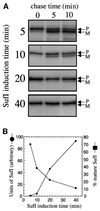
Fig. 2. (A) Saturation of the Tat translocase. MC4100(DE3)arar carrying pET-SufI was grown in M9 minimal media to A600 = 0.4. SufI expression was induced by the addition of IPTG to 1 mM. At 5, 10, 20 and 40 min post-induction, 1.4 ml samples were removed, pulse-labeled for 1 min with [35S]methionine and chased with methionine (500 µg/ml). Aliquots (400 µl) were removed following 0, 5 and 10 min of chase, mixed with 133 µl of 50% TCA, and precipitated proteins were analyzed by Ni-NTA adsorption, SDS–PAGE and fluorography. (B) Quantitation of pre-SufI processing and SufI protein. The percentages of SufI in the mature form were determined for the 10 min chase. For each SufI induction time, an aliquot of the sample chased for 10 min was analyzed by SufI immunoblot analysis using antibody directed against the His6 epitope tag. The protein content for each sample was normalized to the 5 min induction time (set to 1).
None
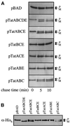
Fig. 3. Overexpression of Tat proteins. For analysis of Tat protein expression, MC4100(DE3)arar carrying either the parental vector pBAD (lane 1) or Tat expression plasmids pTatABCDE (lane 2), pTatABCE (lane 3), pTatBCE (lane 4), pTatACE (lane 5), pTatABE (lane 6) or pTatABC (lane 7) was grown in M9 minimal media to A600 = 0.3. Arabinose was added to 1% and incubation continued for 2 h. An equal number of cells was harvested from each culture, suspended in SDS–PAGE sample buffer, sonicated for 10 s, heated to 37°C for 10 min, and analyzed by SDS–PAGE [High-Tris (Brundage et al., 1990) for TatA and E, 15% for TatB, C and SecY] and immunoblot analysis with affinity-purified antibodies directed against TatA, B, C, E or SecY.
None
Fig. 4. (A) Overexpression of Tat proteins relieves the saturation of Tat translocase. MC4100(DE3)arar carrying pSU-SufI and either the parental vector pBAD or Tat expression plasmids was grown in M9 minimal media lacking methionine to A600 = 0.3. IPTG (1 mM final) and arabinose (1% final) were added and incubation was continued for 45 min. Samples (1.4 ml) were pulse-labeled with [35S]methionine, chased with methionine (500 mg/ml) for 0, 5 and 10 min, and processed for Ni-NTA adsorption, SDS–PAGE and fluorography. (B) Immunoblot analysis of SufI protein. An aliquot from the 10 min chase from each of the samples in (A) was analyzed for SufI protein by immunoblotting using antibody directed against the His6 epitope tag.
None
Fig. 5. (A) SufI post-translational translocation. MC4100(DE3)arar carrying pSU-SufI and either pBAD (lanes 1–4) or pTatABC (lanes 5–8) was grown in M9 minimal media to A600 = 0.4. Samples (0.9 ml) were pulse-labeled with [35S]methionine for 1 min, followed by the addition of methionine (500 mg/ml) and 1% arabinose (lanes 2, 4, 6 and 8). Samples were incubated at 37°C for either 10 min (lanes 1 and 2 and 6 and 7) or 30 min (lanes 3 and 4 and 7 and 8) and 400 µl aliquots were processed for Ni-NTA adsorption, SDS–PAGE and fluorography. (B) Reversal of a CCCP block restores post-translational SufI translocation. MC4100(DE3)arar carrying pET-SufI was grown in M9 minimal media to A600 = 0.4. SufI expression was induced for 5 min with IPTG and samples were then treated with either DMSO (5%) (lanes 1 and 2), DMSO pre-mixed with β-mercaptoethanol (1 mM final) (lane 3), CCCP [100 µM (final) from a 2 mM stock in DMSO] (lanes 4 and 5) or CCCP pre-mixed with β-mercaptoethanol (lane 6) for 1 min, pulse-labeled with [35S]methionine for 1 min and chased with methionine (500 µg/ml). Immediately following the addition of unlabeled methionine, β-mercaptoethanol (1 mM final) was added (lanes 2 and 5). Samples were incubated at 37°C for 10 min, transferred to ice and processed for Ni-NTA adsorption, SDS–PAGE and fluoro graphy. Owing to the decreased incorporation of [35S]methionine in the presence of CCCP, lanes 4 and 5 were exposed longer to achieve comparable signals.
None
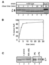
Fig. 6. (AIn vitro translocation of SufI. Complete translocation reactions (50 µl): lanes 3–7 and 9 contained 5 µl of 10 × TL buffer, 300 µg/ml IMVs [either wt (lane 3) or Tat-overexpressed (Tat+, lanes 4–9)], 200 µg/ml lipid-free BSA, 2 mM ATP and 5 mM NADH. The reaction in lane 2 lacked IMVs and lane 8 lacked ATP and had 1 U of potato apyrase. Samples were incubated for 3 min at 37°C and DMSO (1% final) (lane 5) or CCCP (100 µM final from a 10 mM stock in DMSO) (lane 6) was added, followed by 20 µl of 35S-labeled SufI (lanes 1–8) or SufI(RR to KK) (lane 9). Samples were incubated for an additional 60 min at 37°C (lanes 1–6 and 8 and 9) or on ice (lane 7). Reactions were transferred to ice and digested with proteinase K (1 mg/ml) for 15 min. Membranes were sedimented by centrifugation (10 min at 100 000 g), suspended in 100 µl of TL buffer, and protein was precipitated by the addition of TCA (12.5% final) and incubation on ice for 30 min. Precipitated protein was collected by centrifugation (at 14 000 g for 10 min at 4°C), mixed with 1 ml of acetone and sedimented, suspended in 40 µl SDS–PAGE sample buffer, heated at 95°C for 5 min, and analyzed by SDS–PAGE and fluorography. Lane 1 represents 0.5% of the SufI substrate in each translocation reaction. (B) Translocation assays were performed as described in (A), except that membranes were sedimented by centrifugation prior to digestion with proteinase K. During proteinase K digestion, Triton X-100 (1%) was added to the reaction in lane 2. (C) TatABC are required for in vitro translocation. Translocation assays were performed as in (A) containing wild-type, pTatABCE-, pTatABC-, pTatBCE-, pTatACE- or pTatABE-overexpression IMVs (lanes 1–6).

References

    1. Bartolome B., Jubete,Y., Martinez,E. and de la Cruz,F. (1991) Construction and properties of a family of pACYC184-derived cloning vectors compatible with pBR322 and its derivatives. Gene, 102, 75–78. - PubMed
    1. Berks B.C. (1996) A common export pathway for proteins binding complex redox cofactors? Mol. Microbiol., 22, 393–404. - PubMed
    1. Berks B.C., Sargent,F. and Palmer,T. (2000a) The Tat protein export family. Mol. Microbiol., 35, 260–274. - PubMed
    1. Berks B.C., Sargent,F., De Leeuw,E., Hinsley,A.P., Stanely,N.R., Jack,R.L., Buchanan,G. and Palmer,T. (2000b) A novel transport system involved in the biogenesis of bacterial electron transfer chains. Biochim. Biophys. Acta, 1459, 325–330. - PubMed
    1. Bogsch E.G., Sargent,F., Stanley,N.R., Berks,B.C., Robinson,C. and Palmer,T. (1998) An essential component of a novel bacterial protein export system with homologues in plastids and mitochondria. J. Biol. Chem., 273, 18003–18006. - PubMed

Publication types

Substances