. 2001 May 15;20(10):2497-507.
doi: 10.1093/emboj/20.10.2497.

Chemokine receptor homo- or heterodimerization activates distinct signaling pathways

Affiliations

Chemokine receptor homo- or heterodimerization activates distinct signaling pathways

M Mellado et al. EMBO J. .

Abstract

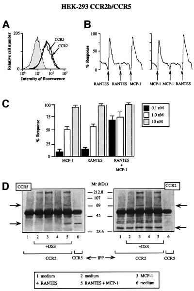
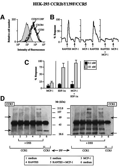
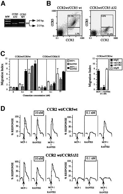
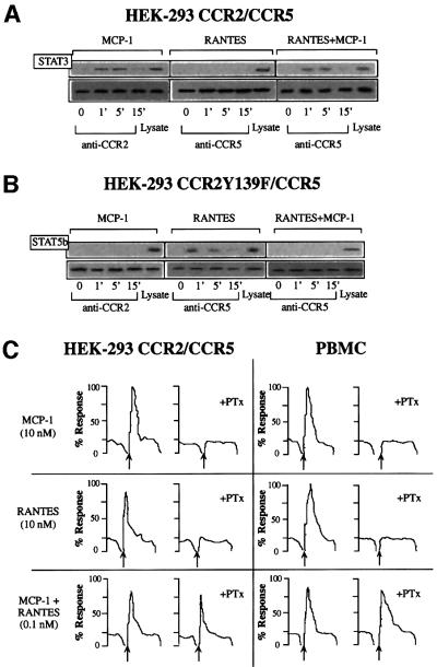
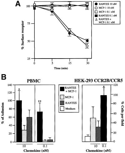
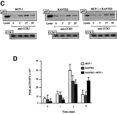
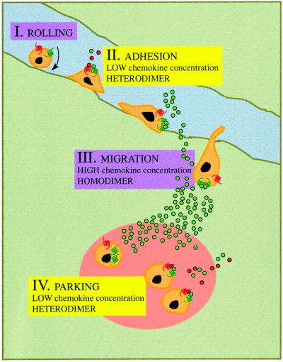
Chemokine receptors of both the CC and CXC families have been demonstrated to undergo a ligand-mediated homodimerization process required for Ca2+ flux and chemotaxis. We show that, in the chemokine response, heterodimerization is also permitted between given receptor pairs, specifically between CCR2 and CCR5. This has functional consequences, as the CCR2 and CCR5 ligands monocyte chemotactic protein-1 (MCP-1) and RANTES (regulated upon activation, normal T cell-expressed and secreted) cooperate to trigger calcium responses at concentrations 10- to 100-fold lower than the threshold for either chemokine alone. Heterodimerization results in recruitment of each receptor-associated signaling complex, but also recruits dissimilar signaling path ways such as G(q/11) association, and delays activation of phosphatidyl inositol 3-kinase. The consequences are a pertussis toxin-resistant Ca2+ flux and trig gering of cell adhesion rather than chemotaxis. These results show the effect of heterodimer formation on increasing the sensitivity and dynamic range of the chemokine response, and may aid in understanding the dynamics of leukocytes at limiting chemokine concentrations in vivo.


Figures

None
Fig. 1. Simultaneous MCP-1 and RANTES co-activation of CCR2- and CCR5-expressing cells increases sensitivity of chemokine responses and promotes their heterodimerization. (A) CCR2b/CCR5 double-transfected HEK-293 cells were incubated with biotin-labeled mAbs against CCR2 and CCR5 or their respective isotype-matched control mAbs, followed by isothiocyanate-labeled streptavidin. (B) Ca2+ mobilization was induced by treatment with 10 nM MCP-1 or 10 nM RANTES in Fluo-3-loaded CCR2/CCR5-co-transfected HEK-293 cells. Results are expressed as a percentage of the chemokine-induced calcium response. Five experiments were performed; the figure depicts one representative experiment. Arrows indicate addition of stimulus. (C) Ca2+ mobilization was determined as in (B), following stimulation with different concentrations of MCP-1 or RANTES as indicated, added separately or simultaneously. Results are expressed as a percentage of the maximum chemokine-induced calcium response. The mean ± SD of four independent experiments is shown. (D) CCR2/CCR5-co-transfected HEK-293 cells were stimulated with chemokines (10 nM for 5 min at 37°C) and, where indicated, cross-linked with 1 mM DSS. Cell lysates were immunoprecipitated with anti-CCR2 antibody, electrophoresed and transferred to nitrocellulose membranes. The western blot was analyzed with anti-CCR5 antibody (left); as a positive control, unstimulated CCR2/CCR5-co-transfected HEK-293 cells were immunoprecipitated with anti-CCR5 antibody (lane 6). The membrane was stripped and reprobed with anti-CCR2 antibody as a control for protein loading (right). Arrows indicate the position to which monomers and dimers migrated.
None
Fig. 2. The mutant CCR2bY139F receptor impairs the response to RANTES but to SDF-1α. (A) CCR2bY139F/CCR5 double-transfected HEK-293 cells were incubated with biotin-labeled mAbs to CCR2, CCR5 or CXCR4 or their respective isotype-matched control mAbs, followed by isothiocyanate-labeled streptavidin. (B) Ca2+ flux was triggered by 10 nM RANTES, 10 nM MCP-1, or a combination of both chemokines (0.1 nM each) as indicated using CCR2bY139F/CCR5-co-transfected HEK-293 cells. Results are expressed as a percentage of the maximum chemokine-induced Ca2+ response. The figure depicts one representative experiment of four performed. (C) Ca2+ mobilization was determined as in Figure 1B, following stimulation of CCR2b Y139F/CCR5-co-transfected HEK-293 cells with different concen trations of MCP-1 or SDF-1α, separately or simultaneously as indicated. Results are expressed as a percentage of the maximum chemokine-induced response. The mean ± SD of three independent experiments is shown. (D) HEK-293 cells co-transfected with the CCR2bY139F and CCR5 receptors were processed as in Figure 1D. Arrows indicate the monomer and the dimer.
None
Fig. 3. Simultaneous MCP-1 and RANTES co-activation of PBMC increases sensitivity of chemokine responses. (A) Amplification of gene fragments corresponding to the CCR5 (245 bp) and CCR5Δ32 (213 bp) with specific primers as described in Materials and methods using genomic DNA from PBMC isolated from CCR5-homozygous and CCR5Δ32-homozygous donors. (B) PBMC from CCR5 wild-type and CCR5Δ32 donors were incubated with anti-CCR2, anti-CCR5 mAbs or their respective isotype-matched control mAbs in the presence of an excess of human immunoglobulins, followed by fluorescein isothiocyanate-labeled anti-mouse IgG and phycoerythrin (PE)-labeled anti-mouse IgM antibodies. The figure also shows the percentage of double-staining cells and single positives. (C) PBMC from CCR5- and CCR5Δ32-homozygous donors were allowed to migrate following stimulation with MCP-1 or RANTES, added separately or simul taneously as indicated. The migration index was calculated as described in Materials and methods. Data represent the mean of quadruplicate determinations, with the SD indicated. Migration of PBMC from CCR5-homozygous donors in response to 0.1 nM MCP-1 plus 0.1 nM RANTES was blocked by pre-treatment of the cells with antibodies against CCR2 and CCR5 (50 µg/ml for 30 min at 37°C). As a control, pre-treatment with isotype-matched antibodies is also shown. (D) Ca2+ flux was triggered by 10 nM or 0.1 nM RANTES, MCP-1, or a combination, using PBMC from CCR5- and CCR5Δ32-homozygous donors. Results are expressed as a percentage of the maximum chemokine-induced calcium response. The figure depicts one representative experiment of four performed.
None
Fig. 4. Heterodimerization of chemokine receptors results in recruitment of specific signaling events. (A) Serum-starved CCR2/CCR5-transfected HEK-293 cells (10 × 106) were used alone or treated with 10 nM MCP-1, RANTES, or a combination of both for the times indicated. Cell lysates were immunoprecipitated with anti-CCR2 (left) or anti-CCR5 antibody (center and right), and western blots were developed with anti-STAT3 antibody. As a control, an unstimulated, unprecipitated transfected HEK-293 cell lysate was analyzed in a western blot with an anti-STAT3 antibody. In each case, CCR2 or CCR5 protein loading was assessed by reprobing membranes with anti-CCR2 or -CCR5 mAb. (B) Serum-starved, CCR2bY139F/CCR5-transfected HEK-293 cells were treated with 10 nM MCP-1, 10 nM RANTES, or a combination of MCP-1 and RANTES (10 nM of each). Cell lysates were immunoprecipitated with anti-CCR2 (left) or anti-CCR5 antibody (center and right) and western blots were developed with anti-STAT5b antibody. Protein loading was controlled for as in (A). (C) Ca2+ mobilization induced by MCP-1 (10 nM), RANTES (10 nM) or MCP-1 plus RANTES (0.1 nM of each) was determined in CCR2- and CCR5-co-transfected HEK-293 cells, and in PBMC from CCR5 wild-type donors untreated or pre-incubated with PTx. The figure depicts one representative experiment of three performed. (D) PBMC from CCR5 wild-type donors were used alone or pre-incubated with PTx, as indicated, and allowed to migrate following stimulation with MCP-1 or RANTES, added separately (10 nM each) or simultaneously (0.1 nM of each) as indicated. The migration index was calculated as described in Materials and methods. Data represent the mean of quadruplicate determinations, with the SD indicated. (E) Serum-starved CCR2- and CCR5-transfected HEK-293 cells (10 × 106) were used alone or treated with 10 nM MCP-1, RANTES, or a combination of both ligands for the times indicated. Cell lysates were immunoprecipitated with anti-CCR2 or anti-CCR5 antibody, and western blots developed with anti-Gq/11 antibody. As a control, an unstimulated, unprecipitated transfected HEK-293 cell lysate was analyzed in a western blot with anti-Gq/11 antibody. As a protein loading control, membranes were reprobed with the immunoprecipitating antibody.
None
Fig. 5. Chemokine receptor heterodimerization promotes specific signaling events but not receptor down-regulation. (A) Serum-starved CCR2/CCR5-transfected HEK-293 cells (10 × 106) were incubated for the times indicated with 0.1 nM and 10 nM RANTES, 0.1 nM and 10 nM MCP-1, or 0.1 nM of both chemokines at 37°C. Surface CCR2 or CCR5 was detected by fluorescence-activated cell sorting (FACS) analysis using biotin-labeled CCR2-03 mAb or CCR5-03 mAb, followed by streptavidin–PE; an isotype-matched mAb was used as control. Results are expressed as the percentage of maximum binding obtained in the absence of chemokines, with the SD indicated. (B) A static adhesion assay was performed using CCR2b- and CCR5-transfected HEK-293 cells (right) or PBMC from CCR5 wild-type donors (left), as described in Materials and methods. Stimuli include MCP-1 (10 nM), RANTES (10 nM), or MCP-1 plus RANTES (0.1 nM of each) for transfected cells, and 10 nM or 0.1 nM of MCP-1, RANTES, or both chemokines together for PBMC, as indicated. Results are expressed as a percentage of the maximum adhesion observed after stimulation with a mixture of RANTES plus MCP-1. The figure shows the mean ± SD of seven independent experiments. *Significantly different (p <0.05); **significantly different (p <0.01) (Student’s t-test). (C) Serum-starved CCR2- and CCR5-transfected HEK-293 cells (10 × 106) were incubated for the times indicated with 10 nM RANTES, 10 nM MCP-1, or 0.1 nM of each of these chemokines at 37°C. Cell lysates were immunoprecipitated with anti-CCR2 or anti-CCR5 antibody as indicated, and western blots were developed with anti-p85 antibodies (upper panel). CCR2 or CCR5 protein loading was controlled for as in Figure 4A (lower panel). (D) Serum-starved CCR2- and CCR5-transfected HEK-293 cells (10 × 106) were incubated for the indicated times with 10 nM RANTES, 10 nM MCP-1, or 0.1 nM of each of these chemokines combined at 37°C. Cell lysates were immunoprecipitated with anti-CCR2 or anti -CCR5 antibody, and an in vitro kinase assay was performed as indicated (Materials and methods). The mean ± SD of three independent experiments is shown.
None
Fig. 6. The physiological role of chemokine receptor dimerization. Leukocytes roll along the blood vessel endothelium (I); exposure to low chemokine concentrations causes the formation of chemokine receptor heterodimers and the cell adheres to the endothelium (II). The inflammatory response produces higher chemokine concentrations, triggering receptor homodimerization; this induces cell migration through the endothelium to inflammation sites (III), where low chemokine concentrations favor heterodimerization, leading the cell to adhere (‘park’) in the tissues (IV).

References

    1. Al-Aoukaty A., Schall,T.J. and Maghazachi,A.A. (1996) Differential coupling of CC chemokine receptors to multiple heterotrimeric G proteins in human interleukin 2-activated natural killer cells. Blood, 87, 4255–4260. - PubMed
    1. Alon U., Surette,M.G., Barkai,N. and Leibler,S. (1999) Robustness in bacterial chemotaxis. Nature, 397, 168–171. - PubMed
    1. Aragay A., Frade,J.M.R., Mellado,M., Serrano,A., Martínez-A.,C. and Mayor,F.,Jr (1998) MCP-1-induced CCR2B receptor desensitization by the G protein-coupled receptor kinase-2. Proc. Natl Acad. Sci. USA, 95, 2985–2990. - PMC - PubMed
    1. Arai H. and Charo,I.F. (1996) Differential regulation of G-protein mediated signaling by chemokine receptors. J. Biol. Chem., 271, 21814–21819. - PubMed
    1. Baggiolini M. (1998) Chemokines and leukocyte traffic. Nature, 392, 565–568. - PubMed

Publication types