. 2001 May 15;20(10):2518-27.
doi: 10.1093/emboj/20.10.2518.

Interactions between the Su(Hw) and Mod(mdg4) proteins required for gypsy insulator function

Affiliations

Interactions between the Su(Hw) and Mod(mdg4) proteins required for gypsy insulator function

D Ghosh et al. EMBO J. .

Abstract

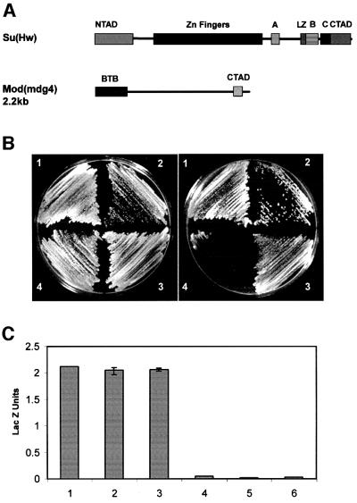
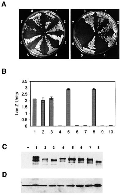
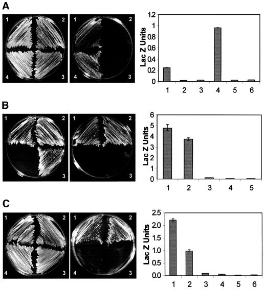
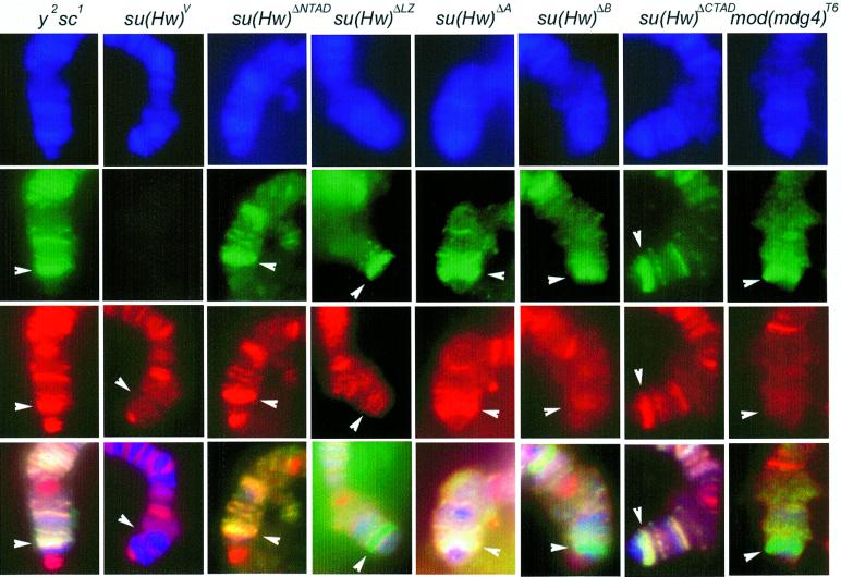
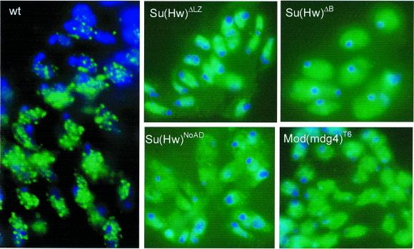
The gypsy insulator is thought to play a role in nuclear organization and the establishment of higher order chromatin domains by bringing together several individual insulator sites to form rosette-like structures in the interphase nucleus. The Su(Hw) and Mod(mdg4) proteins are components of the gypsy insulator required for its effect on enhancer-promoter interactions. Using the yeast two-hybrid system, we show that the Mod(mdg4) protein can form homodimers, which can then interact with Su(Hw). The BTB domain of Mod(mdg4) is involved in homodimerization, whereas the C-terminal region of the protein is involved in interactions with the leucine zipper and adjacent regions of the Su(Hw) protein. Analyses using immunolocalization on polytene chromosomes confirm the involvement of these domains in mediating the interactions between these proteins. Studies using diploid interphase cells further suggest the contribution of these domains to the formation of rosette-like structures in the nucleus. The results provide a biochemical basis for the aggregation of multiple insulator sites and support the role of the gypsy insulator in nuclear organization.


Figures

None
Fig. 1. Su(Hw) and mod(mdg4) interact directly with each other. (A) Schematic map of the Su(Hw) and Mod(mdg4) proteins showing the various domains described in the text. NTAD, N-terminal acidic domain; CTAD, C-terminal acidic domain; LZ, leucine zipper; A, B and C denote regions defined by homology among different Drosophila species (Harrison et al., 1993). BTB represents the BTB/POZ domain of mod(mdg4) and CTAD the C-terminal acidic domain. (B) Growth of yeast strain pJ694A expressing different Su(Hw) and/or Mod(mdg4) proteins on non-selective (left) or selective (right) media for the reporter genes used in the yeast two-hybrid assays. The numbers on the plates denote the following: 1, yeast expressing Su(Hw)-GAL4BD and Mod(mdg4)-GAL4AD; 2, yeast expressing Mod(mdg4)-GAL4BD and Su(Hw)-GAL4AD; 3, yeast expressing Mod(mdg4)-GAL4BD and Mod(mdg4)-GAL4AD; 4, yeast expressing Su(Hw)-GAL4BD and Su(Hw)-GAL4AD. (C) β-galactosidase activity, expressed as Miller units, in extracts of yeast strains carrying combinations of full-length Su(Hw) and Mod(mdg4) protein. Numbers 1–4 correspond to yeast strains described above. Numbers 5 and 6 correspond to yeast expressing Su(Hw) and Mod(mdg4) alone.
None
Fig. 2. Interactions between Su(Hw) and Mod(mdg4) are mediated by the leucine zipper and regions B and C of Su(Hw). (A) Growth of yeast strains carrying full-length Mod(mdg4) fused to GAL4BD and different deletion constructs of Su(Hw) fused to GAL4AD on non-selective (left) or selective (right) media. The numbers on the plates denote yeast expressing Mod(mdg4)–GAL4AD and either full-length or deletions of Su(Hw) fused to GAL4BD. 1, full-length Su(Hw); 2, Su(Hw)ΔNTAD; 3, Su(Hw)ΔCTAD; 4, Su(Hw)ΔLZ; 5, Su(Hw)ΔA; 6, Su(Hw)ΔB; 7, Su(Hw)ΔC; 8, Su(Hw)ΔNoAD2. (B) β-galactosidase activities, expressed as Miller units, corresponding to strains carrying combinations of full-length Mod(mdg4) and different deletion constructs of Su(Hw). Numbers denote the same strains described above. Numbers 9 and 10 denote yeast expressing Su(Hw) and Mod(mdg4) alone. (C) Western analyses of yeast extracts carrying different deletions of Su(Hw). The panel shows the expression of Su(Hw) detected with polyclonal anti-Su(Hw) antibody. Numbers are as in the previous panels. The ‘–’ symbol represents extracts from yeast cells not expressing Su(Hw) or Mod(mdg4). (D) Same western as in (C), after the filter was stripped and re-probed with a porin monoclonal antibody. The presence of mitochondrial porin was used as a loading control.
None
Fig. 3. Domains of Su(Hw) and Mod(mdg4) necessary and sufficient for interactions. The photographs on the left show growth of yeast strains carrying deletions of Mod(mdg4) and full-length Su(Hw) or full-length Mod(mdg4) on media containing (left panel) or lacking (right panel) histidine and adenine. The graphs on the right show β-galactosidase activity, expressed as Miller units, in extracts from strains carrying the same combinations of Mod(mdg4) and Su(Hw). For each graph, numbers 1–4 are as on the photographs to the left, and numbers 5 and 6 correspond to yeast expressing Su(Hw) and Mod(mdg4) alone. (A) The numbers on the plates denote yeast expressing the following constructs: 1, yeast expressing Su(Hw)–GAL4BD and Mod(modg4)ΔBTB–GAL4AD; 2, Mod(mdg4)–GAL4BD and Mod(mdg4)ΔBTB–GAL4AD; 3, Su(Hw)–GAL4BD and Mod(mdg4)ΔCTAD–GAL4AD; 4, Mod(mdg4)–GAL4BD and Mod(mdg4)ΔCTAD–GAL4AD. (B) The numbers on the plates denote the following: 1, yeast transformed with Mod(mdg4)–GAL4BD and LZ + B + C + CTAD–GAL4AD; 2, Mod(mdg4)–GAL4BD and LZ + B + C–GAL4AD; 3, BTB–GAL4BD and LZ + B + C–GAL4AD. Numbers 4 and 5 in the graph correspond to yeast expressing Su(Hw) and Mod(mdg4) alone. (C) The numbers on the plates correspond to yeast transformed with: 1, BTB–GAL4BD and Mod(mdg4)–GAL4AD; 2, BTB–GAL4BD and BTB–GAL4AD; 3, BTB–GAL4BD and Mod(mdg4)ΔBTB–GAL4AD; 4, BTB–GAL4AD and Su(Hw)–GAL4AD.
None
Fig. 4. Interactions between Su(Hw) and Mod(mdg4) in Drosophila polytene chromosomes. Localization of Su(Hw) (green) and Mod(mdg4) (red) on polytene chromosomes; 4′,6-diamidine-2-phenylindole (DAPI) stains DNA and is indicated in blue. The lower panels represent the overlap of the three individual images. All strains carry a gypsy-induced mutation in the yellow gene and they are either wild type for su(Hw) and mod(mdg4) (y2) or carry different mutations in these genes as indicated at the top of the various panels. Su(Hw) and Mod(mdg4) proteins were detected using FITC- and Texas red-conjugated secondary antibodies, respectively. Sites where Su(Hw) is present alone should be labeled in green, whereas sites for Mod(mdg4) should be marked in red; sites where both proteins are present appear yellow. The location of the yellow locus is indicated by a white arrowhead.
None
Fig. 5. Subnuclear localization of Su(Hw) in nuclei. Diploid cells in interphase were obtained from imaginal discs of Drosophila wild type (wt) or strains carrying different mutations in Su(Hw) and Mod(mdg4) as indicated in the panels. Distribution of Su(Hw) detected with FITC-conjugated secondary antibody (green). DNA was stained with DAPI and is represented in blue.

References

    1. Ahmad K.F., Engel,C.K. and Prive,G.G. (1998) Crystal structure of the BTB domain from PLZF. Proc. Natl Acad. Sci. USA, 95, 12123–12128. - PMC - PubMed
    1. Bardwell V.J. and Treisman,R. (1994) The POZ domain: a conserved protein–protein interaction motif. Genes Dev., 8, 1664–1677. - PubMed
    1. Bell A.C. and Felsenfeld,G. (1999) Stopped at the border: boundaries and insulators. Curr. Opin. Genet. Dev., 9, 191–198. - PubMed
    1. Bell A.C., West,A.G. and Felsenfeld,G. (1999) The protein CTCF is required for the enhancer blocking activity of vertebrate insulators. Cell, 98, 387–396. - PubMed
    1. Bell A.C., West,A.G. and Felsenfeld,G. (2001) Insulators and boundaries: Versatile regulatory elements in the eukaryotic genome. Science, 291, 447–450. - PubMed

Publication types