Review
. 2001 May 15;533(Pt 1):57-63.
doi: 10.1111/j.1469-7793.2001.0057b.x.

Sacral spinal interneurones and the control of urinary bladder and urethral striated sphincter muscle function

Affiliations
Review

Sacral spinal interneurones and the control of urinary bladder and urethral striated sphincter muscle function

S J Shefchyk. J Physiol. .

Abstract

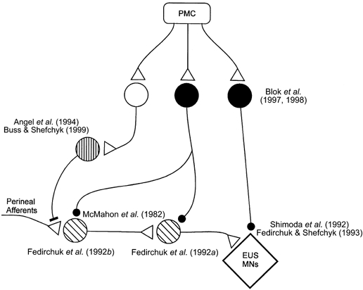
Normally, during bladder filling (continence) and expulsion (micturition) there is a reciprocity between the pattern of activity in the urinary bladder sacral parasympathetic efferents and the somatic motoneurones innervating the striated external urethral sphincter muscle. The co-ordination of this pattern of reciprocal activity appears to be determined by excitatory and inhibitory actions of a variety of segmental afferents and descending systems with sacral spinal actions. These actions may in part be mediated through lower lumbar and sacral excitatory and inhibitory spinal interneurones. Over the past 30 years, both neuroanatomical and electrophysiological approaches have been used to reveal an ever-increasing richness in the neuronal network in the lower spinal cord related to the bladder and striated external urethral sphincter muscle. The purpose of this review is to present an overview of the identified excitatory and inhibitory spinal interneurones hypothesized to be involved in the central networks controlling the sacral bladder parasympathetic preganglionic neurones and striated urethral sphincter motoneurones during continence and micturition.


Figures

Figure 1
Figure 1. Schematic diagram of the basic spino-bulbo-spinal micturition reflex
The afferent signal from the bladder is carried by the pelvic nerve and is relayed up the spinal cord to the pontine micturition centre (PMC), which functions as a switch between continence and micturition. Descending signals from the brainstem then produce the excitation of the bladder parasympathetic neurones carried in the pelvic nerve and a decrease in pudendal nerve efferent activity to the striated urethral sphincter muscle. Triangular terminals represent excitation while filled circles represent inhibition. Arrows indicate pathways that may not be direct. PGNs, parasympathetic preganglionic neurones; EUS MNs, external urethral sphincter motoneurones.
Figure 2
Figure 2. Schematic diagram of the neurones thought to be involved in the control of the sacral bladder parasympathetic preganglionic neurones
Triangular terminals represent excitatory synapses while filled circles represent inhibitory synapses. Arrows represent connections that are implied but may not be direct. For ease of illustration neurones are distinguished by different shading of the cell bodies and some polysynaptic pathways may be represented by only one or two interneurones where more may exist. Citations appearing near the cells refer to the studies described in the text. PGNs, parasympathetic preganglionic neurones; PMC, pontine micturition centre.
Figure 3
Figure 3. Schematic diagram of the neurones thought to be involved in the control of the ventral horn motoneurones innervating the striated external urethral sphincter muscle
The format is the same as in Fig. 2 with the addition of a filled bar terminal representing synapses mediating presynaptic inhibition of primary afferents. For ease of illustration the interneurones are distinguished by different shading of the cell bodies; these do not correspond to the interneurones represented in Fig. 2. EUS MNs, external urethral sphincter motoneurones; PMC, pontine micturition centre.

Similar articles

Cited by

References

    1. Alvarez FJ. Anatomical basis for presynaptic inhibition of primary sensory fibers. In: Rudomin P, Romo R, Mendell LM, editors. Presynaptic Inhibition and Neural Control. New York: Oxford University Press; 1998. pp. 13–49.
    1. Angel MJ, Fyda D, Mccrea DA, Shefchyk SJ. Primary afferent depolarization of cat pudendal afferents during micturition and segmental afferent stimulation. Journal of Physiology. 1994;479:451–461. - PMC - PubMed
    1. Araki I. Inhibitory postsynaptic currents and the effects of GABA on visually identified sacral parasympathetic preganglionic neurons in neonatal rats. Journal of Neurophysiology. 1994;72:2903–2910. - PubMed
    1. Araki I, de groat WC. Unitary excitatory synaptic currents in preganglionic neurons mediated by two distinct groups of interneurons in neonatal rat sacral parasympathetic nucleus. Journal of Neurophysiology. 1996;76:215–226. - PubMed
    1. Araki I, de groat WC. Developmental synaptic depression underlying reorganization of visceral reflex pathways in the spinal cord. Journal of Neuroscience. 1997;17:8402–8407. - PMC - PubMed

Publication types

LinkOut - more resources