Review
. 2001 May 15;533(Pt 1):99-109.
doi: 10.1111/j.1469-7793.2001.0099b.x.

Neural prostheses

Affiliations
Review

Neural prostheses

A Prochazka et al. J Physiol. .

Abstract

Assuming that neural regeneration after spinal cord injury (SCI) will eventually become a clinical reality, functional recovery will probably remain incomplete. Assistive devices will therefore continue to play an important role in rehabilitation. Neural prostheses (NPs) are assistive devices that restore functions lost as a result of neural damage. NPs electrically stimulate nerves and are either external or implanted devices. Surface stimulators for muscle exercise are now commonplace in rehabilitation clinics and many homes. Regarding implantable NPs, since 1963 over 40 000 have been implanted to restore hearing, bladder control and respiration. Epidural spinal cord stimulators and deep brain stimulators are routinely implanted to control pain, spasticity, tremor and rigidity. Implantable NPs have also been developed to restore limb movements using electrodes tunnelled under the skin to muscles and nerves. Spinal cord microstimulation (SC[mu]stim) is under study as an alternative way of restoring movement and bladder control. Improvement in bladder and bowel function is a high priority for many SCI people. Sacral root stimulation to elicit bladder contraction is the current NP approach, but this usually requires dorsal rhizotomies to reduce reflex contractions of the external urethral sphincter. It is possible that the spinal centres coordinating the bladder-sphincter synergy could be activated with SC[mu]stim. Given the large and growing number of NPs in use or development, it is surprising how little is known about their long-term interactions with the nervous system. Physiological research will play an important role in elucidating the mechanisms underlying these interactions.


Figures
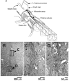
Figure 1
Figure 1. Microwire implantation technique and histology
A, 6-12 microwires were implanted in each side of the spinal cord with their tips targeting the ventral horn (1.7-2.1 mm from the midline, 3-4.2 mm deep). The microwires were spaced 2-3 mm apart along the rostral-caudal length of the lumbar enlargement. B, visible electrode track in a 6 μm cross-section of the spinal cord, stained with haematoxylin and eosin and embedded in paraffin. Boxed regions are shown at higher magnification in C and D. C, close-up of the microwire track. Note that the tissue capsule is only about 50 μm in diameter (microwire occupying 30 μm) and does not include any signs of an enduring inflammatory response (i.e. no astrocytes or lymphocytes are present). D, detail of the area around the microwire tip. No signs of electrically induced neural damage are seen and nearby motoneurone seems intact.
Figure 2
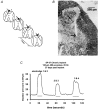
Figure 2. Scheme for chronic microstimulation in the feline sacral spinal cord
A, the arrays of 4 individual iridium microelectrodes were implanted at rostro-caudal intervals of approximately 1 mm, and directed towards the intermediolateral cell column and the preganglionic parasympathetic nucleus. B, site of the tip of one of the microelectrodes (arrow), close to the PPN (1 μm section of epoxy-embedded spinal cord, stained with Toluidine Blue and Azure II). C, hydrostatic pressure within the urinary bladder induced by stimulating with various pairs of microelectrodes. The stimulus pulses were applied to the 2 electrodes in an interleaved manner. (Modified from Woodford et al. 1996, with permission.)
Figure 3
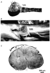
Figure 3. Integrated microelectrode array for chronic implantation in the sacral spinal cord
A, the array of 6 iridium microelectrodes, each approximately 50 μm in diameter, extends from an epoxy button whose underside is contoured to approximate the curvature of the dorsal spinal cord. B, an array in situ in a cat's sacral spinal cord, 35 days after implantation. C, histological section through the cat's sacral spinal cord, and parallel to one of the electrode tracks. The tip of the track (arrowhead) is slightly dorsal to the centre of its target, the intermediolateral cell column (asterisk). (D. B. McCreery, A. S. Lossinsky, W. F. Agnew & L. A. Bullara, unpublished observations.)

Similar articles

Cited by

References

    1. Accornero N, Bini G, Lenzi GL, Manfredi M. Selective activation of peripheral nerve fibre groups of different diameter by triangular-shaped stimulus pulses. Journal of Physiology. 1977;273:539–560. - PMC - PubMed
    1. Al-Majed AA, Neumann CM, Brushart TM, Gordon T. Brief electrical stimulation promotes the speed and accuracy of motor axonal regeneration. Journal of Neuroscience. 2000;20:2602–2608. - PMC - PubMed
    1. Benabid A, Koudsie A, Benazzouz A, Fraix V, Ashraf A, Le bas JF, Chabardes S, Pollak P. Subthalamic stimulation for Parkinson's disease. Archives of Medical Research. 2000;31:282–289. - PubMed
    1. Benabid AL, Pollak P, Gervason C, Hoffmann D, Gao DM, Hommel M, Perret JE, de rougemont J. Long-term suppression of tremor by chronic stimulation of the ventral intermediate thalamic nucleus. Lancet. 1991;337:403–406. - PubMed
    1. Blok BFM, Holstege G. The central nervous system control of micturition in cats and humans. Behavioural Brain Research. 1998;92:119–125. - PubMed

Publication types