. 2001 May;126(1):47-68.
doi: 10.1104/pp.126.1.47.

The arabidopsis cell plate-associated dynamin-like protein, ADL1Ap, is required for multiple stages of plant growth and development

Affiliations

The arabidopsis cell plate-associated dynamin-like protein, ADL1Ap, is required for multiple stages of plant growth and development

B H Kang et al. Plant Physiol. 2001 May.

Abstract

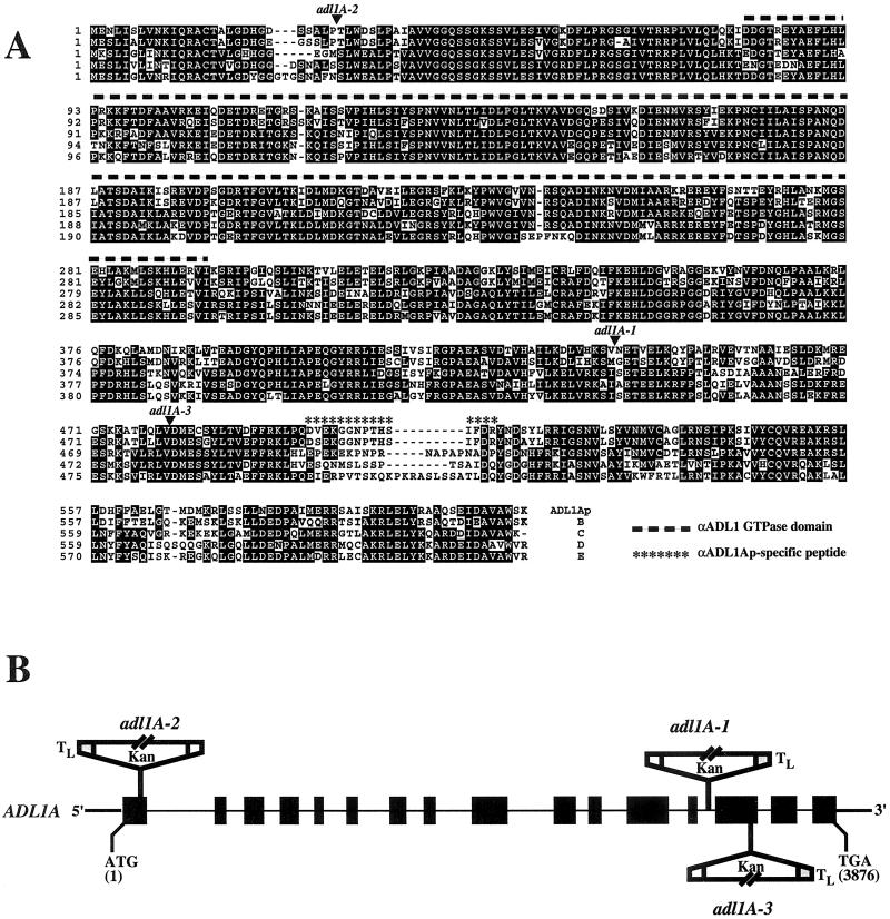
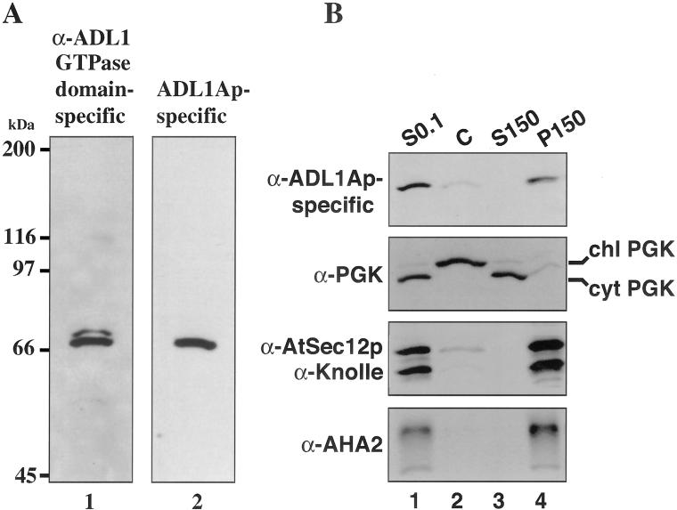
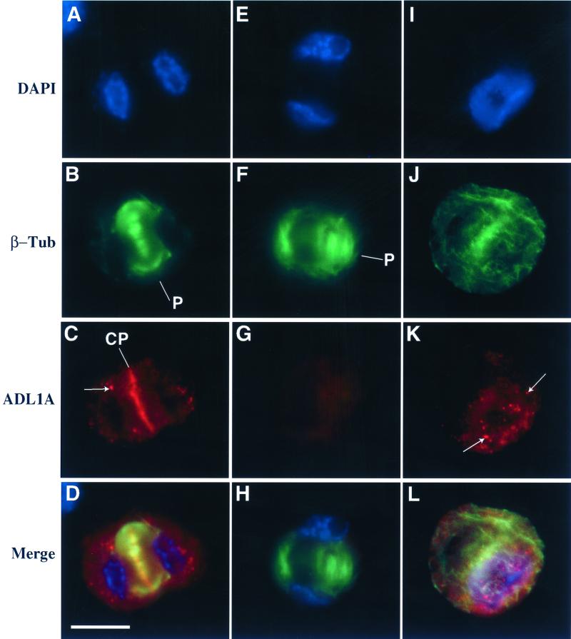
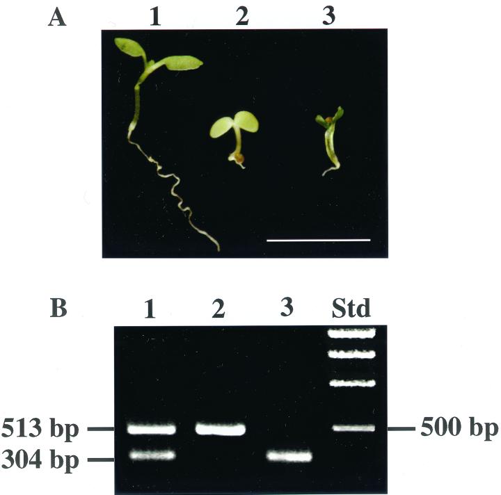
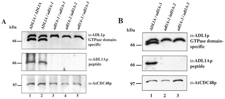
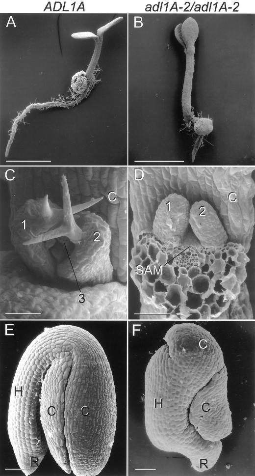
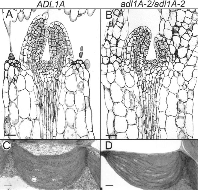
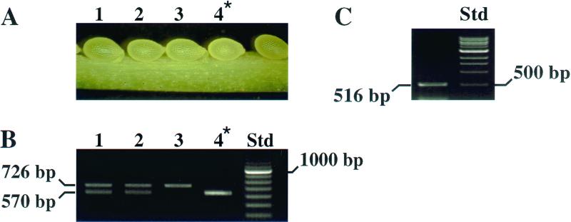
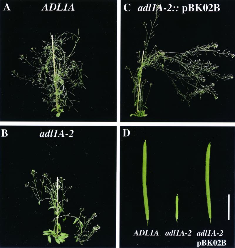
Dynamin and dynamin-like proteins are GTP-binding proteins involved in vesicle trafficking. In soybean, a 68-kD dynamin-like protein called phragmoplastin has been shown to be associated with the cell plate in dividing cells (Gu and Verma, 1996). Five ADL1 genes encoding dynamin-like proteins related to phragmoplastin have been identified in the completed Arabidopsis genome. Here we report that ADL1Ap is associated with punctate subcellular structures and with the cell plate in dividing cells. To assess the function of ADL1Ap we utilized a reverse genetic approach to isolate three separate Arabidopsis mutant lines containing T-DNA insertions in ADL1A. Homozygous adl1A seeds were shriveled and mutant seedlings arrested soon after germination, producing only two leaf primordia and severely stunted roots. Immunoblotting revealed that ADL1Ap expression was not detectable in the mutants. Despite the loss of ADL1Ap, the mutants did not display any defects in cytokinesis, and growth of the mutant seedlings could be rescued in tissue culture by the addition of sucrose. Although these sucrose-rescued plants displayed normal vegetative growth and flowered, they set very few seeds. Thus, ADL1Ap is critical for several stages of plant development, including embryogenesis, seedling development, and reproduction. We discuss the putative role of ADL1Ap in vesicular trafficking, cytokinesis, and other aspects of plant growth.


Figures

Figure 1
Figure 1
Arabidopsis 68-kD dynamin-like protein open reading frames. A, Alignment of the deduced amino acid sequences of the protein encoded by ADL1A, ADL1Ap (GenBank accession no. G2129608), and four other Arabidopsis approximately 68-kD dynamin-like protein open reading frames (GenBank accession nos. G6850867 [B], G8778229 [C], G3341679 [D], and G7076772 [E]). The alignment was generated using the Multiple Alignment Program (Smith et al., 1996). Identical and conserved amino acid residues are outlined in black and gray, respectively. Dashes depict gaps included to maximize sequence similarity. The 15-amino acid non-conserved hydrophilic region of ADL1Ap that was used to generate ADL1Ap-specific antibodies is overlined with asterisks. The 168-amino acid segment used to generate the general anti-ADL1p GTPase domain-specific antibody is overlined with bold dashes. The arrowheads indicate the positions at which the adl1A mutant proteins would likely be truncated. B, Schematic representation of ADL1A gene disruptions. The exon/intron structure of ADL1A is shown to scale, with black boxes representing exons. The positions of the translation initiation and termination codons are signified by ATG and TGA, respectively. The positions and orientations of T-DNA inserts with left border sequences (TL; not drawn to scale) are indicated. Kan, T-DNA neomycin phosphotransferase selectable gene marker.
Figure 2
Figure 2
Immunodectection and characterization of ADL1Ap. A, Analysis of ADL1Ap-specific antibodies. Twenty micrograms of post-nuclear supernatant (S0.1) prepared from suspension-cultured protoplasts was fractionated on 7.5% (w/v) SDS-polyacrylamide gels and was analyzed by immunoblotting. Equal loading was confirmed by Ponceau S staining. Immunoblots were probed with (1) affinity-purified antibodies directed against the ADL1p GTPase domain, or (2) affinity-purified antibodies specific for ADL1Ap and visualized by enhanced chemiluminescence. B, Subcellular distribution of ADL1Ap. Chloroplasts (C), total microsomal membranes (P150), and membrane-free cytosol (S150) were isolated from a 100g Arabidopsis protoplast post-nuclear supernatant (S0.1). Fifteen micrograms of protein from each fraction was separated by SDS-PAGE and was analyzed by immunoblotting using ADL1Ap-specific antibodies. The fractionation of ADL1Ap was compared with the distribution of various soluble and membrane subcellular markers, including cytosolic PGK (cytPGK) and chloroplast-associated PGK (chlPGK), AtSec12p (endoplasmic reticulum), AHA2 (plasma membrane), and Knolle (cell plate).
Figure 3
Figure 3
Immunolocalization of ADL1Ap in Arabidopsis suspension-cultured cells. Protoplasts from a 3-d culture of Arabidopsis cells were double immunolabeled with antibodies directed against β-tubulin (β-Tub) to visualize cortical and phragmoplast microtubules (B, F, and J) and either affinity-purified ADL1Ap-specfic (C and K) or preimmune sera (G). Localization of the nuclear material was revealed by staining with 4′,6′-diamidino-2-phenylindole (DAPI; A, E, and I). Electronically merged images of cells in cytokinesis (A–C) and (E–G) as demonstrated by the presence of two nuclei and interphase (I–K) are shown in D, H, and L, respectively. Bar = 50 μm, P, Phragmoplast; CP, cell plate. Arrows indicate ADL1Ap-positive subcellular structures.
Figure 4
Figure 4
Genotype analysis of adl1A–2 seedlings. A, Five-day old wild-type kanr (1), wild-type kans (2), and mutant seedlings (3) grown on germination media in continuous light (bar = 5.0 mm). B, Total DNA was prepared from individual 5-d-old seedlings and was analyzed by PCR using a mixture of three primers specific to ADL1A and the left T-DNA border (TL; see “Materials and Methods”). PCR amplification of DNA from plants homozygous for the wild-type ADL1A gene yielded only a single approximately 0.5-kb product (lane 2), whereas genomic DNA from the heterozygous adl1::T-DNA-tagged plant (lane 1) yielded the wild-type 0.5-kb product and the approximately 0.3-kb T-DNA-tagged product. Homozygous adl1A-2 seedlings (lane 3) yielded only a single approximately 0.3-kb PCR product.
Figure 5
Figure 5
Analysis of ADL1Ap expression in wild-type and adl1A seedlings. A, Total protein was prepared from 5-d wild-type (lane 1), heterozygous (lane 2), and homozygous adl1A seedlings (lanes 3–5) grown on germination medium and analyzed by SDS-PAGE and immunoblotting. B, Immunoblot analysis of leaf total protein from sugar-treated heterozygous adl1A-3 (lane 1) and “sugar-rescued” homozygous adl1A-2 and adl1A-3 plants (lanes 2 and 3). The immunoblots were probed as indicated using affinity-purified ADL1p GTPase domain, ADL1Ap-specific antibodies, and AtCdc48p antibodies. AtCdc48p was used as a loading control. In addition, equal loading was confirmed by Ponceau S staining.
Figure 6
Figure 6
Embryonic and seedling phenotype of adl1A mutants. Scanning electron micrographs of Arabidopsis 3-d-old wild-type seedling (A) and 5-d-old adl1A-2/adl1A-2 seedlings (B). C, Wild-type shoot apex with three visible leaf primordia, labeled 1 through 3, above the cotyledons; D, adl1A-2/adl1A-2 shoot apex; one cotyledon has been removed to visualize the apex. Two rosette leaf primordia, labeled 1 and 2, are visible on the flanks of the mutant shoot apical meristem. E, Wild-type mature embryo curved such that the hypocotyl-root-axis is parallel to the two cotyledons; F, adl1A-2/adl1A-2 mature embryo with two cotyledons wrapped around the hypocotyl. A twist occurs in the lower portion of the mutant hypocotyl. A and B, Bars = 1 mm. C through F, Bars = 50 μm. C, Cotyledon; H, hypocotyl; R, radicle.
Figure 7
Figure 7
Shoot apical meristem structure of adl1A mutants. Light micrographs of median longitudinal sections through shoot apices and first two rosette leaf primordia of wild-type (A) and mutant adl1A-2/adl1A-2 (B) seedlings. The tunica corpus apical organization of the mutant (B) is the same as that of the wild-type (A). Transmission electron micrographs of chloroplasts from upper palisade tissue of wild-type (C) and adl1A-2/adl1A-2 (D) cotyledons. A and B, Bars = 25 μm. C and D, Bars = 0.5 μm.
Figure 8
Figure 8
ADL1Ap functions during late embryogenesis. A, Portion of an immature silique from a heterozygous adl1A-1 plant, approximately 10 to 12 d after flowering. Twenty-five percent of the seeds in a green silique are pale green (asterisks). B, PCR analysis of the genotype of individual embryos isolated from developing seeds shown in A. Genomic DNA from individual embryos was analyzed by PCR (see “Materials and Methods”); DNA from plants homozygous for the wild-type ADL1A gene (lane 3) yields only a single approximately 0.7-kb product when the PCR amplification is performed with the TL + 3′ + 5′ primers. In contrast, PCR amplification of genomic DNA from the heterozygous adl1::T-DNA-tagged plant (lanes 1 and 2) yields the wild-type approximately 0.7-kb product and the approximately 0.6-kb T-DNA-tagged product. Homozygous adl1A embryos (lane 4, asterisk) yield only a single approximately 0.6-kb PCR product. C, RT-PCR analysis of ADL1A expression in wild-type isolated embryos. cDNA was synthesized from total RNA isolated from mature-“walking-stick stage” embryos (Goldberg et al., 1994). To confirm that the PCR-products were derived from reverse transcribed RNA and not from contaminating genomic DNA, the ADL1A -specific primers were designed to amplify a approximately 600-bp fragment of ADL1A containing an approximately 100-bp intron and were then sequenced.
Figure 9
Figure 9
Phenotype of flowering rescued ald1A plants and siliques. Flowering wild-type (A), homozygous “Suc-rescued” adl1A-2 (B), and transgenic homozygous adl1A-2: :pBK02B (C) plants containing an extragenic copy of ADL1A. D, Siliques from wild-type, “Suc-rescued” adl1A-2, and adl1A-2::pBK02B plants; bar = 5.0 mm.
Figure 10
Figure 10
Molecular analysis of homozygous and complemented adl1A-2 plants. PCR analysis of homozygous adl1A-2 transformed with the binary transformation vector pBK02B. A, Two oligonucleotide pairs, I =(SB7 + SB60) and II = (SB7 + SB59), were used for PCR to distinguish wild-type, heterozygous, and homozygous adl1A-2::pBK02B transgenic plants. Oligonucleotides SB7, SB60, and SB59 are specific to the 5′ end of ADL1A, pBK02B, and to the 3′ genomic DNA sequence flanking ADL1A, which was not included in the pBK02B, respectively. Primer pair I is specific to pBK02B and II is specific to the endogenous wild-type copy of ADL1A. B, PCR amplification of genomic DNA from untransformed wild-type ADL1A/ADL1A plants (lanes 1 and 2), transgenic ADL1A/adl1A-2: :pBK02B plants (lanes 3 and 4), transgenic homozygous adl1A-2::pBK02B plants (lanes 5 and 6), and purified pBK02B plasmid DNA (lanes 7 and 8). PCR analysis of DNA from homozygous adl1A-2::pBK02B plants confirms the presence of the complementing ADL1A genomic copy and confirms that the plants are homozygous for the T-DNA insertion in ADL1A. Cross-hatched box, pBK02B vector DNA; speckled box, P1 MJC20 DNA flanking the 15-kB XhoI/KpnI restriction fragment containing ADL1A; gray triangle, ADL1A upstream regulatory sequences; gray square, ADL1A 3′ untranslated sequence.

References

    1. Assaad FF, Mayer U, Wanner G, Jürgens G. The KEULE gene is involved in cytokinesis in Arabidopsis. Mol Gen Genet. 1996;253:267–277. - PubMed
    1. Axelos M, Curie C, Mazzolini L, Bardet C, Lescure B. A protocol for transient gene expression in Arabidopsis thaliana protoplasts isolated from cell suspension cultures. Plant Physiol Biochem. 1992;30:123–128.
    1. Bar-Peled M, Raikhel NV. Characterization of AtSec12 and AtSAR1 proteins likely involved in ER and Golgi traffic. Plant Physiol. 1997;114:315–324. - PMC - PubMed
    1. Baumlein H, Nagy I, Villarroel R, Inzé D, Wobus U. Cis-analysis of a seed protein gene promoter: the conservative RY repeat CATGCATG within the legumin box is essential for tissue-specific expression of a legumin gene. Plant J. 1992;2:233–239. - PubMed
    1. Bednarek SY, Wilkins TA, Dombrowski JE, Raikhel NV. A carboxyl-terminal propeptide is necessary for proper sorting of barley lectin to vacuoles of tobacco. Plant Cell. 1990;2:1145–1155. - PMC - PubMed

Publication types