. 2001 May;126(1):210-21.
doi: 10.1104/pp.126.1.210.

Altered middle lamella homogalacturonan and disrupted deposition of (1-->5)-alpha-L-arabinan in the pericarp of Cnr, a ripening mutant of tomato

Affiliations

Altered middle lamella homogalacturonan and disrupted deposition of (1-->5)-alpha-L-arabinan in the pericarp of Cnr, a ripening mutant of tomato

C Orfila et al. Plant Physiol. 2001 May.

Abstract

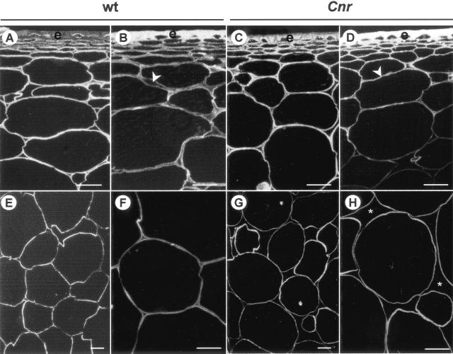
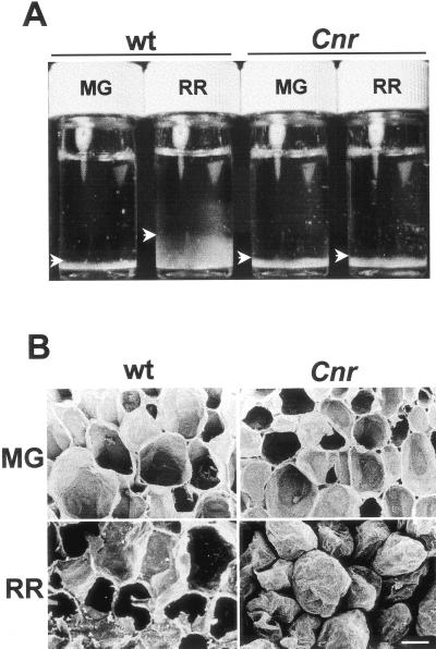
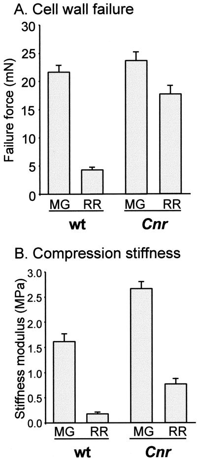
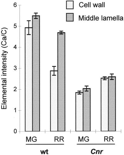
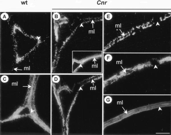
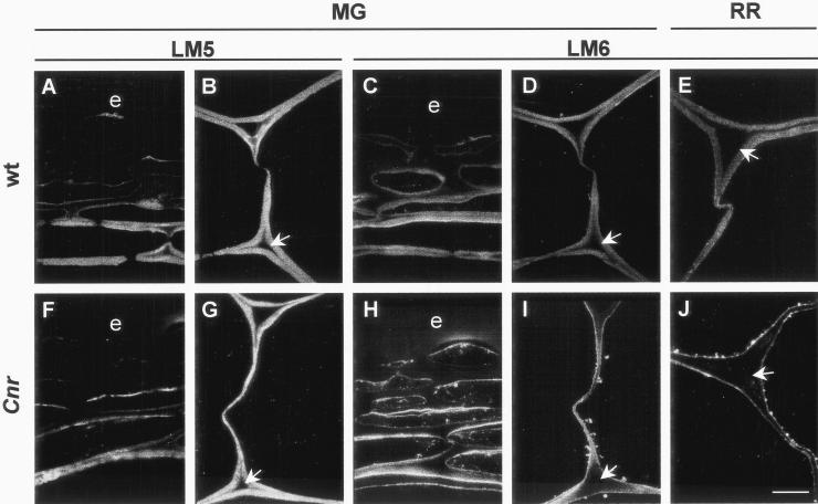
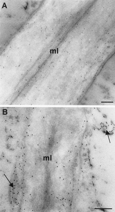
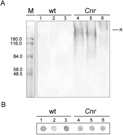
Cnr (colorless non-ripening) is a pleiotropic tomato (Lycopersicon esculentum) fruit ripening mutant with altered tissue properties including weaker cell-to-cell contacts in the pericarp (A.J. Thompson, M. Tor, C.S. Barry, J. Vrebalov, C. Orfila, M.C. Jarvis, J.J. Giovannoni, D. Grierson, G.B. Seymour [1999] Plant Physiol 120: 383-390). Whereas the genetic basis of the Cnr mutation is being identified by molecular analyses, here we report the identification of cell biological factors underlying the Cnr texture phenotype. In comparison with wild type, ripe-stage Cnr fruits have stronger, non-swollen cell walls (CW) throughout the pericarp and extensive intercellular space in the inner pericarp. Using electron energy loss spectroscopy imaging of calcium-binding capacity and anti-homogalacturonan (HG) antibody probes (PAM1 and JIM5) we demonstrate that maturation processes involving middle lamella HG are altered in Cnr fruit, resulting in the absence or a low level of HG-/calcium-based cell adhesion. We also demonstrate that the deposition of (1-->5)-alpha-L-arabinan is disrupted in Cnr pericarp CW and that this disruption occurs prior to fruit ripening. The relationship between the disruption of (1-->5)-alpha-L-arabinan deposition in pericarp CW and the Cnr phenotype is discussed.


Figures

Figure 1
Figure 1
Calcofluor White staining of cellulose in resin-embedded sections of pericarp. A, Outer pericarp of MG WT fruit. B, Outer pericarp of RR WT fruit. Arrowhead indicates expanded ML. C, Outer pericarp of MG Cnr fruit. D, Outer pericarp of RR Cnr fruit. Arrowhead indicates compact cell walls. E and F, Regions of the inner pericarp of RR WT fruit. G and H, Regions of the inner pericarp of RR Cnr fruit. Asterisks indicate large intercellular spaces. e, Epidermis. Bars = 200 μm.
Figure 2
Figure 2
CW properties. A, CW swelling in vitro. Equivalent dry weights of isolated CWM from WT and Cnr pericarp at MG and RR stages were suspended in water and allowed to hydrate at room temperature for 8 h with gentle agitation then allowed to settle. CWM is seen at the base of each vial (arrowheads). B, Scanning electron micrographs of fracture surfaces of fruit pericarp of WT and Cnr at MG and RR stages. RR Cnr pericarp cells separate and do not rupture. Bar = 200 μm.
Figure 3
Figure 3
Mechanical analyses of pericarp material. A, Force required for CW failure in WT and Cnr MG and RR fruit. Error bars indicate se of the mean (n = 6). B, Compression stiffness moduli for WT and Cnr MG and RR fruit. Error bars indicate se of the mean (n = 6).
Figure 4
Figure 4
Calcium-binding capacity for WT and Cnr pericarp tissue at MG and RR developmental stages represented as the Ca/C elemental intensity ratios. Error bars indicate ses of the mean.
Figure 5
Figure 5
Immunolabeling of pectic HG components in regions of intercellular spaces in outer pericarp. A, Intercellular space of RR WT pericarp. The arrow indicates the ML. Arrowhead indicates abundant PAM1 labeling at corner of intercellular space. B, Intercellular space RR Cnr pericarp. Arrow indicates ML without PAM1 binding. Arrowhead indicates absence of PAM1 epitope from corner of space. Inset shows a serially related section of RR Cnr pericarp labeled with JIM5. C, Intercellular space of RR WT pericarp de-esterified before PAM1 probing. Arrow indicates abundance of PAM1 epitope at the ML. D, Intercellular space of RR Cnr pericarp de-esterified before PAM1 probing. Arrow indicates absence of PAM1 epitope from ML and corners of space. E and F, Region of CW away from intercellular space. The PAM1 epitope is absent from regions of the CW (arrowhead), even after de-esterification (F). G, Serially related section to E and F probed with JIM5 shows epitope throughout the CW and abundantly at the ML. In each case, arrow indicates ML. A through D, Arrowhead indicates corner of intercellular space. Bar = 5 μm for all micrographs.
Figure 6
Figure 6
Immunofluorescent labeling of (1→4)-β-galactan and (1→5)-α-arabinan epitopes in tomato pericarp shows disrupted deposition of (1→5)-α-arabinan in Cnr fruit. A and B, The LM5 (1→4)-β-galactan epitope in resin-embedded sections of outer region of the pericarp (A) and inner pericarp CW (B) of MG WT fruit. C and D, The LM6 (1→5)-α-arabinan epitope in resin-embedded sections of outer region of the pericarp (C) and inner pericarp CW (D) of MG WT fruit. E, The LM6 (1→5)-α-arabinan epitope in resin-embedded sections of inner pericarp CW of RR WT fruit. F and G, The LM5 (1→4)-β-galactan epitope in resin-embedded sections of outer region of the pericarp (F) and inner pericarp CW (G) of MG Cnr fruit. H and I, The LM6 (1→5)-α-arabinan epitope in resin-embedded sections of outer region of the pericarp (H) and inner pericarp CW (I) of MG Cnr fruit. Throughout the pericarp the LM6 epitope occurs in vesicles located in the peripheral cytoplasm and in the inner region of the CW. J, The disruption of the LM6 (1→5)-α-arabinan epitope deposition and its absence from CW is particularly clear in the inner pericarp of RR WT fruit. Arrows indicate in each case the CW lining intercellular space. e, Epidermis. Bar = 10 μm.
Figure 7
Figure 7
Immunogold electron microscopy of the (1→5)-α-arabinan epitope in MG tomato pericarp CW. A, In WT pericarp CW the LM6 (1→5)-α-arabinan epitope was evenly distributed. B, In Cnr pericarp CW the LM6 (1→5)-α-arabinan epitope occurred most abundantly at inner regions of the CW and was also associated with vesicle-like structures in the cytoplasm and at the plasma membrane (indicated by arrows). Bars = 200 nm.
Figure 8
Figure 8
Immunochemistry of whole-cell extracts with anti-(1→5)-α-arabinan probe, LM6. A, Immunoblotting on nitrocellulose with LM6 of material separated by SDS-PAGE. Material (10 μg of protein per lane) from WT MG fruit (lanes 1–3) and Cnr MG fruit (lanes 4–6) were prepared in sample buffer immediately (lanes 1 and 4) or after incubation in Tris-buffer (lanes 2 and 5) or incubation in Tris-buffer plus Pronase E (lanes 3 and 6). Significant amounts of LM6-reactive, Pronase E-sensitive material entered the gel from Cnr, but not WT material. R indicates top of resolving gel. M shows Mr markers. B, Immunodot assay on nitrocellulose of material (0.4 μg of protein per dot) analyzed in A showing abundance of LM6 epitope in all samples.

References

    1. Albersheim P, Darvill AG, O'Neill MA, Schols HA, Voragen AGJ. An hypothesis: the same six polysaccharides are components of the primary cell walls of all higher plants. In: Visser J, Voragen AGJ, editors. Pectins and Pectinases. Amsterdam: Elsevier Science; 1996. pp. 47–55.
    1. Blakeney AB, Harris PJ, Henry RJ, Stone BA. A simple and rapid preparation of alditol acetates for monosaccharide analysis. Carbohydr Res. 1983;113:291–299.
    1. Blumenkrantz N, Absoe-Hansen G. New method for quantitative determination of uronic acids. Anal Biochem. 1973;54:484–489. - PubMed
    1. Brady CJ. Fruit ripening. Annu Rev Plant Physiol. 1987;38:155–178.
    1. Carey AT, Holt K, Picard S, Wilde R, Tucker GA, Bird CR, Schuch W, Seymour GB. Tomato exo-(1–4)-β-d-galactanase: isolation, changes during ripening in normal and mutant tomato fruit, and characterization of a related cDNA clone. Plant Physiol. 1995;108:1099–1107. - PMC - PubMed

Publication types

MeSH terms

LinkOut - more resources