. 2001 Jul 20;276(29):26784-91.
doi: 10.1074/jbc.M104038200. Epub 2001 May 14.

The binding of the circumsporozoite protein to cell surface heparan sulfate proteoglycans is required for plasmodium sporozoite attachment to target cells

Affiliations

The binding of the circumsporozoite protein to cell surface heparan sulfate proteoglycans is required for plasmodium sporozoite attachment to target cells

C Pinzon-Ortiz et al. J Biol Chem. .

Abstract

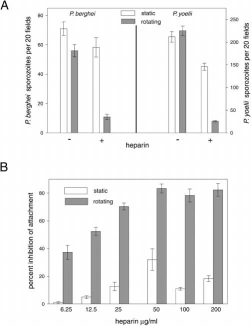
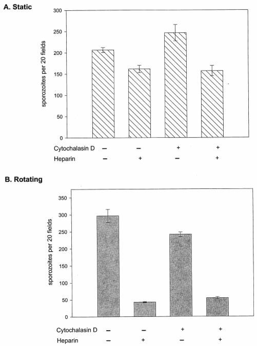
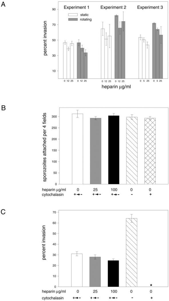
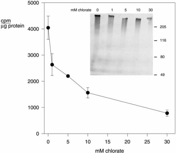
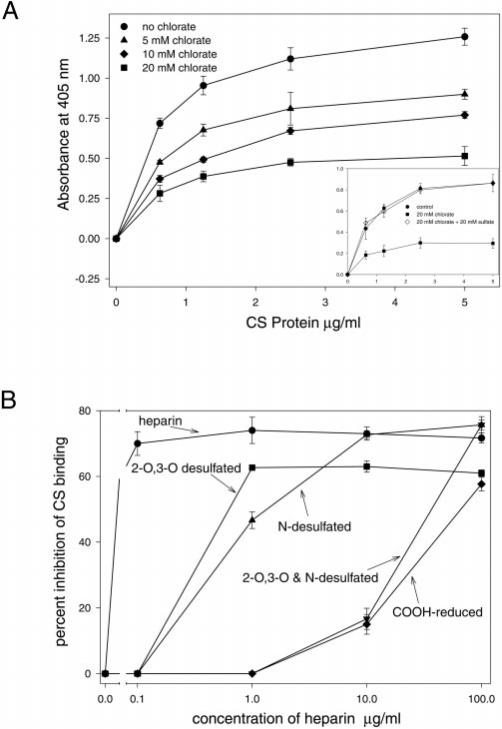
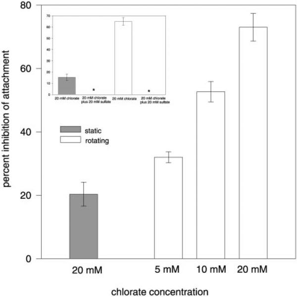
The major surface protein of malaria sporozoites, the circumsporozoite protein, binds to heparan sulfate proteoglycans on the surface of hepatocytes. It has been proposed that this binding event is responsible for the rapid and specific localization of sporozoites to the liver after their injection into the skin by an infected anopheline mosquito. Previous in vitro studies performed under static conditions have failed to demonstrate a significant role for heparan sulfate proteoglycans during sporozoite invasion of cells. We performed sporozoite attachment and invasion assays under more dynamic conditions and found a dramatic decrease in sporozoite attachment to cells in the presence of heparin. In contrast to its effect on attachment, heparin does not appear to have an effect on sporozoite invasion of cells. When substituted heparins were used as competitive inhibitors of sporozoite attachment, we found that sulfation of the glycosaminoglycan chains at both the N- and O-positions was important for sporozoite adhesion to cells. We conclude that the binding of the circumsporozoite protein to hepatic heparan sulfate proteoglycans is likely to function during sporozoite attachment in the liver and that this adhesion event depends on the sulfated glycosaminoglycan chains of the proteoglycans.


Figures

Fig. 1
Fig. 1. Heparin is a better inhibitor of sporozoite attachment under conditions that mimic flow
Sporozoites were preincubated ± 25 μg/ml of heparin (A) or the indicated concentration of heparin (B) for 15 min on ice and then plated on cells in the continued presence of heparin. After 1 h at 37 °C under static or rotating conditions, unattached sporozoites were removed by washing, and the attached sporozoites were visualized with the appropriate monoclonal antibody (mAb 3D11 for P. berghei and mAb NYS1 for P. yoelii) and goat anti-mouse Ig conjugated to FITC. Each point was plated in triplicate and shown are the means with standard deviations. A, attachment of P. berghei and P. yoelii sporozoites to cells under static and rotating conditions. B, dose-dependent inhibition of P. berghei sporozoite attachment. Percent inhibition was calculated using the mean number of sporozoites attached in the absence of heparin under static or rotating conditions.
Fig. 2
Fig. 2. Heparin inhibition of sporozoite attachment to HepG2 cells in a parallel plate flow chamber
Sporozoites were preincubated ± 25 μg/ml of heparin for 15 min on ice, infused into the inlet tubing of the flow chamber, and then perfused over the cells at controlled flow rates. The wall shear stress was calculated as a function of flow rate. The perfusion time was inversely proportional to the flow rate so that for each point an equal number of sporozoites were allowed to perfuse over the cell monolayer. Nonadherent sporozoites were washed off with medium infused at 0.05 ml/min (resulting in a shear force of 0.25 dynes/cm2) for 6 min. The cells were fixed, and sporozoites were stained as outlined previously. For each point, the entire area covered by the flow chamber was divided into 2, and ≥100 fields per region were counted. Percent inhibition was calculated using the mean number of sporozoites attached in the absence of heparin with the same flow rate.
Fig. 3
Fig. 3. Sporozoite attachment to HepG2 cells in the presence of heparin and cytochalasin D
P. berghei sporozoites were preincubated with or without 1 μm cytochalasin D ± 25 μg/ml heparin and then plated on cells (in the continued presence of these compounds). After 1 h at 37 °C under static (A) or rotating (B) conditions, unattached sporozoites were removed, and the cells were stained with a double-staining procedure that allows the differentiation of intracellular from extracellular sporozoites. Each point was plated in triplicate and shown are the means with standard deviations of the total number of sporozoites attached to the cells. Invasion data for this experiment is not shown; however, in the presence of cytochalasin D there was no invasion, and in the absence of cytochalasin D, ~40% of attached sporozoites were found intracellularly regardless of whether heparin was present or not.
Fig. 4
Fig. 4. Heparin does not inhibit sporozoite invasion of HepG2 cells
A, sporozoite invasion in the presence of heparin under static and rotating conditions. P. berghei sporozoites were preincubated in medium ± heparin (25 μg/ml) and then plated on cells in the presence of heparin. After 1 h at 37 °C under static or rotating conditions, unattached sporozoites were removed, and the cells were stained with a double-staining procedure that allows the differentiation of intracellular from extracellular sporozoites. The percentage of sporozoites that invaded the cells was calculated using the following equation: ((total parasites – extracellular parasites)/total parasites) × 100. Each point was plated in triplicate and shown are the means with standard deviations. Because of the variation in invasion efficiency among different batches of sporozoites, we show results from three separate experiments. B and C, sporozoite invasion after recovery from cytochalasin treatment. P. berghei sporozoites were preincubated in medium with 1 μm cytochalasin, added to cells, and allowed to adhere under static conditions in the continued presence of cytochalasin. After 30 min, the cytochalasin-containing medium and any unattached sporozoites were removed; medium containing the indicated concentrations of heparin was added, and sporozoites were allowed to invade cells in the presence of heparin. White bars, no heparin; gray bars, 25 μg/ml heparin; black bars, 100 μg/ml heparin. Controls included sporozoites preincubated and added to cells in medium without cytochalasin or heparin (slanted line bars) and sporozoites preincubated and maintained in medium with cytochalasin for the entire experiment (cross-hatched bar). Sporozoites were allowed to invade cells for 1 h and then the cells were double-stained so that intracellular and extracellular sporozoites could be distinguished. The total number of sporozoites attached for each experimental condition (B) and the percentage of attached sporozoites that were found intracellularly (C) are shown. None of the sporozoites that were in the continuous presence of cytochalasin were found intracellularly (the asterisk in the graph indicates that the data were collected but the error bar cannot be seen since it is 0). Each point was plated in triplicate and shown are the means with standard deviations.
Fig. 5
Fig. 5. Chlorate inhibits sulfate incorporation into proteoglycans of HepG2 cells
HepG2 cells were plated in 6-well plates in medium with the indicated concentration of chlorate. After 12 h, the medium was changed to low sulfate medium with the indicated concentrations of chlorate and [35S]sodium sulfate. 12 h later the cells were washed, lysed, and total cell-associated counts/min were measured in a β-counter. Each point was performed in triplicate and shown are the means with standard deviations. Inset, lysates from chlorate-treated cells were loaded onto a 5% SDS-polyacrylamide gel that was then subjected to autoradiography. Equivalent amounts of protein were loaded onto each lane. Most of the labeled material migrated as a broad high molecular weight smear, typical of proteoglycans.
Fig. 6
Fig. 6
A, CS binding to chlorate-treated HepG2 cells. Cells were plated in 96-well plates and grown in low sulfate medium with the indicated concentrations of chlorate. After 18–24 h, they were fixed, blocked, and incubated with increasing amounts of CS protein. CS binding was revealed with mAb 2A10 specific for the CS repeats, followed by anti-mouse Ig conjugated to horseradish peroxidase and peroxidase substrate. The CS binding curves for cells grown in 20 mm chlorate (squares), 10 mm chlorate (diamonds), 5 mm chlorate (triangles), and no added chlorate (circles) are shown. Each point was assayed in triplicate, and the means with standard deviations are shown. Inset shows CS binding curves for a control experiment where cells were grown in low sulfate medium with 20 mm chlorate (squares), 20 mm chlorate plus 20 mm magnesium sulfate (open diamonds), or no chlorate (circles). B, inhibition of CS binding to HepG2 cells with modified heparins. 5 μg/ml CS was preincubated with the indicated concentrations of each heparin for 30 min at 37 °C. These solutions were then added to paraformaldehyde-fixed HepG2 cells for 1 h at 37 °C; the cells were washed, and bound CS was determined using iodinated mAb 2A10, specific for the CS repeats. Shown is the percent inhibition of binding of CS to HepG2 cells in the presence of inhibitor compared with results obtained in the absence of inhibitor. Inhibitors were heparin (circles), 2-O,3-O-desulfated heparin (squares), N-desulfated heparin (triangles), 2-O,3-O- and N-desulfated heparin (inverted triangles), and carboxy-reduced heparin (diamonds). Each inhibitor concentration was assayed in triplicate, and the means with standard deviations are shown.
Fig. 7
Fig. 7. Inhibition of sporozoite attachment to chlorate-treated HepG2 cells
HepG2 cells were plated in labtek chamber slides, and 2 days later the medium was replaced with low sulfate medium containing the indicated concentrations of chlorate. After 24 h, the chlorate-containing medium was removed, and P. berghei sporozoites in DMEM/FCS were added to each well. After 1 h at 37 °C under static (gray bars) or rotating conditions (white bars), unattached sporozoites were removed; the cells were fixed, and the attached sporozoites were stained. Shown is the percent inhibition of sporozoite attachment to cells in the presence of chlorate compared with the mean number of sporozoites attached in the absence of chlorate (under static or rotating conditions). Each point was plated in triplicate and shown are the means with standard deviations. Inset shows sporozoite attachment, under static and rotating conditions, to cells that had been incubated (as above) in medium with no chlorate, 20 mm chlorate, and 20 mm chlorate plus 20 mm magnesium sulfate. There was no inhibition of sporozoite attachment to cells in 20 mm chlorate plus 20 mm magnesium sulfate under both static and rotating conditions (asterisks indicate that the data were collected, but the error bar cannot be seen since it is 0). Each point was plated in triplicate and shown are the means with standard deviations.
Fig. 8
Fig. 8. Sporozoite attachment to HepG2 cells under rotating conditions in the presence of modified heparins
P. berghei sporozoites were preincubated for 15 min on ice in medium alone or with 25 mg/ml heparin (white bar), 2-O,3-O-desulfated heparin (gray bar), N-desulfated heparin (black bar), 2-O,3-O- and N-desulfated heparin (cross-hatched bar), and carboxy-reduced heparin (diagonally lined bar) and then added to HepG2 cells in labtek chamber slides. Sporozoite incubation with cells was under rotating conditions and in the continued presence of the inhibitor. After 1 h, unattached sporozoites were removed; the cells were fixed, and the attached sporozoites were stained and counted. Each point was plated in triplicate wells. Percent inhibition of sporozoite attachment was calculated using the mean number of sporozoites attached in the absence of heparin. This experiment was performed three times and shown are the pooled results from all three experiments.

References

    1. Sinnis P, Sim BKL. Trends Microbiol. 1997;5:52–58. - PMC - PubMed
    1. Cerami C, Frevert U, Sinnis P, Takacs B, Nussenzweig V. J. Exp. Med. 1994;179:695–701. - PMC - PubMed
    1. Sinnis P, Willnow TE, Briones MRS, Herz J, Nussenzweig V. J. Exp. Med. 1996;184:945–954. - PMC - PubMed
    1. Pancake SJ, Holt GH, Mellouk S, Hoffman SL. J. Cell Biol. 1992;6:1351–1357. - PMC - PubMed
    1. Renia L, Miltgen F, Charoenvit Y, Ponnudurai T, Verhave JP, Collins WE, Mazier D. J. Immunol. Methods. 1988;112:201–205. - PubMed

Publication types

MeSH terms

Substances

LinkOut - more resources