. 2001 May 14;153(4):637-48.
doi: 10.1083/jcb.153.4.637.

The drosophila protein asp is involved in microtubule organization during spindle formation and cytokinesis

Affiliations

The drosophila protein asp is involved in microtubule organization during spindle formation and cytokinesis

J G Wakefield et al. J Cell Biol. .

Abstract

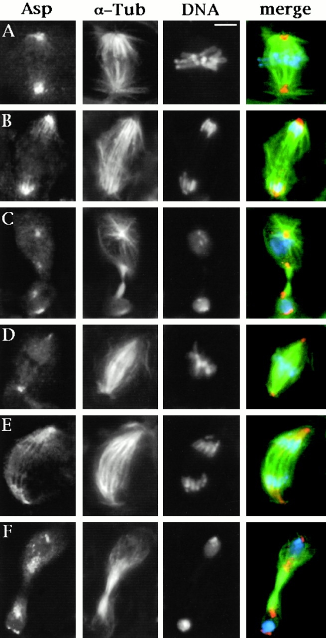
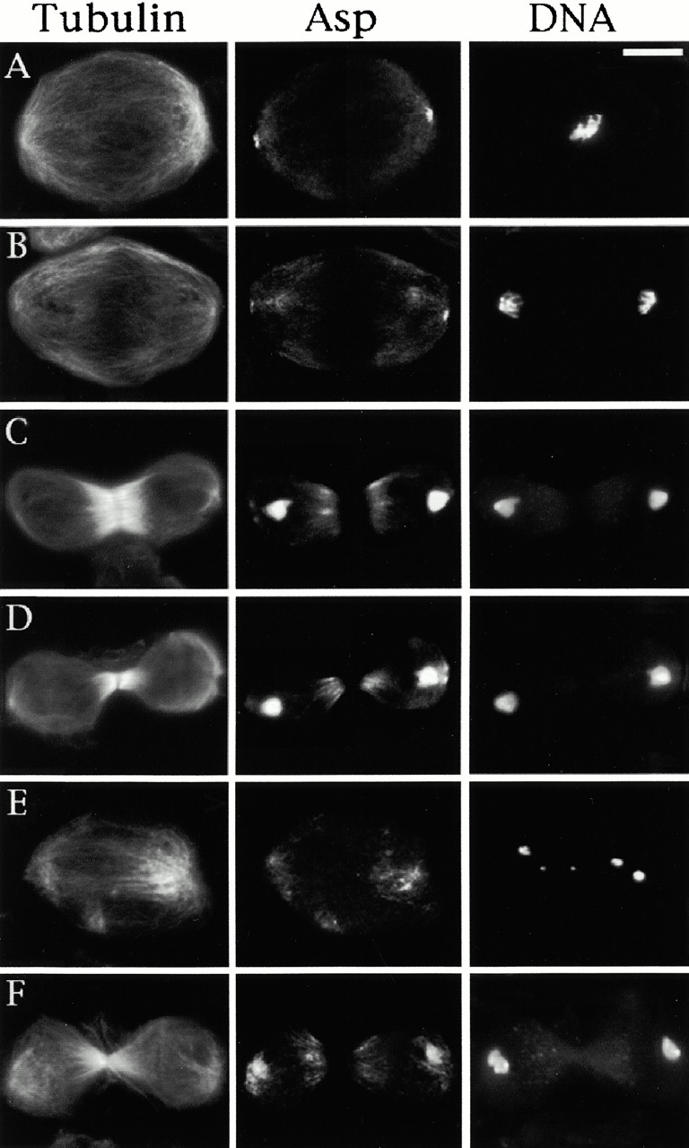
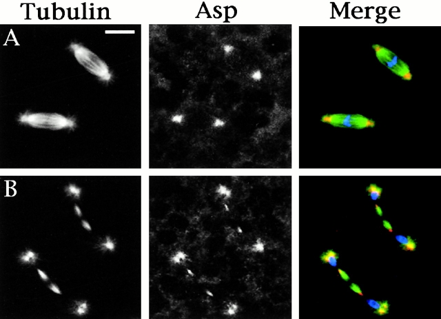
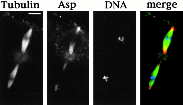
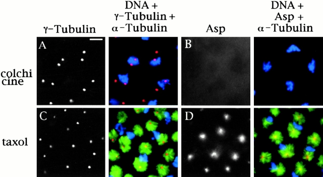
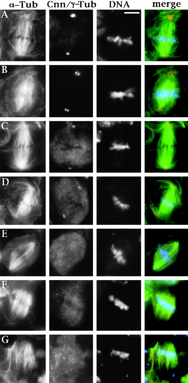
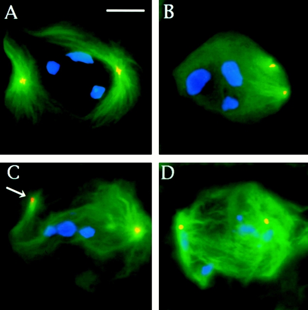
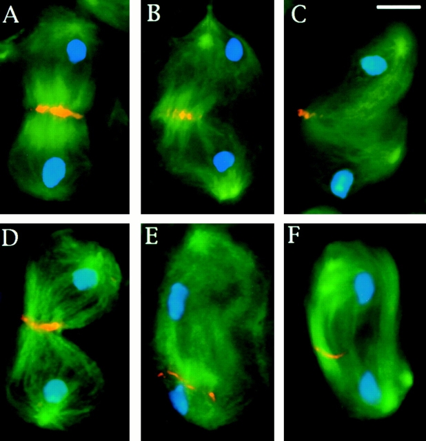
Abnormal spindle (Asp) is a 220-kD microtubule-associated protein from Drosophila that has been suggested to be involved in microtubule nucleation from the centrosome. Here, we show that Asp is enriched at the poles of meiotic and mitotic spindles and localizes to the minus ends of central spindle microtubules. Localization to these structures is independent of a functional centrosome. Moreover, colchicine treatment disrupts Asp localization to the centrosome, indicating that Asp is not an integral centrosomal protein. In both meiotic and mitotic divisions of asp mutants, microtubule nucleation occurs from the centrosome, and gamma-tubulin localizes correctly. However, spindle pole focusing and organization are severely affected. By examining cells that carry mutations both in asp and in asterless, a gene required for centrosome function, we have determined the role of Asp in the absence of centrosomes. Phenotypic analysis of these double mutants shows that Asp is required for the aggregation of microtubules into focused spindle poles, reinforcing the conclusion that its function at the spindle poles is independent of any putative role in microtubule nucleation. Our data also suggest that Asp has a role in the formation of the central spindle. The inability of asp mutants to correctly organize the central spindle leads to disruption of the contractile ring machinery and failure in cytokinesis.


Figures

Figure 3
Figure 3
Asp localization in neuroblasts of wild-type and asl larval brains. Cells were stained for α-tubulin, Asp, and DNA (by DAPI). In the merged images, DNA is colored in blue, tubulin in green, and Asp in orange. (A–C) Wild-type neuroblasts; (D–F) asl neuroblasts; (A and D) metaphases; (B and E) anaphases; (C and F) telophases. Note that both wild-type and asl neuroblasts divide asymmetrically, giving rise to two daughter cells of different sizes (C and F). For details on unequal neuroblast division see Giansanti et al. 2001. In metaphase and anaphase figures, Asp is localized at the spindle poles, wheras in telophases (C and F) it accumulates at both the poles and the extremities of the central spindle. Note that wild-type and asl neuroblasts exhibit similar patterns of Asp accumulation. Bar, 5 μm.
Figure 1
Figure 1
Asp localization during the first meiotic division of wild-type, asp, and asl males. Cells were fixed according to Giansanti et al. 1999 (described in Materials and Methods) and stained for α-tubulin, Asp, and DNA (by DAPI). (A–D) Wild-type meiotic cells. (A) Metaphase; (B) early anaphase; (C) late anaphase; (D) telophase. Note that in metaphase (A) figures, Asp is localized at the spindle poles. In telophase cells (D), Asp predominantly accumulates at the extremities of the central spindle but not in the central spindle midzone. (E) An asl anaphase-like figure showing Asp staining at the spindle poles. (F) An asl telophase with a well-organized central spindle showing a regular Asp immunostaining at its extremities. Bar, 10 μm.
Figure 2
Figure 2
Asp localization in cells of wild-type Drosophila embryos. In the merged images, DNA is colored in blue, tubulin in green, and Asp in orange. (A) Metaphases showing Asp localization at the spindle poles; (B) telophases showing Asp accumulation at the minus ends of central spindle microtubules. Bar, 10 μm.
Figure 4
Figure 4
Asp localization in cells undergoing the second meiotic division in wild-type Drosophila oocytes. In the merged image, DNA is colored in blue, tubulin in green, and Asp in orange. Note that Asp accumulates at the poles of both meiotic spindles. Bar, 10 μm.
Figure 5
Figure 5
Localization of Asp in embryos treated with colchicine or taxol. Embryos were incubated in 5 mM colchicine or 25 μM taxol for 20 min before fixation (described in Materials and Methods). Embryos from the same batch were stained with antibodies to either γ- and α-tubulin (A and C) or Asp and α-tubulin (B and D) and with propidium iodide to show the mitotic chromatin (A–D). In the merged images, chromatin is colored in blue, tubulin in green, and Asp or γ-tubulin in red/orange. (A and B) 5 mM colchicine. (A) α-Tubulin staining is diffuse, suggesting all microtubules have been depolymerised. γ-Tubulin, as an integral centrosomal protein, is tightly focused at the centrosome. (B) α-Tubulin is not detectable, and Asp does not show any centrosomal localization. (C and D) 25 μM taxol. (C) Microtubules are stabilized and form astral arrays surrounding each centrosome. γ-Tubulin is found only at the centrosomes. (D) Asp staining is found throughout the area corresponding to the minus ends of the microtubules, and it is not focused at the centrosome. Bar, 10 μm.
Figure 6
Figure 6
Spindle pole organization in asp neuroblast metaphases. Cells were stained for α-tubulin, DNA, and either centrosomin (Cnn; A and B) or γ-tubulin (C–G). In the merged images, chromatin is colored in blue, α-tubulin in green, and Cnn or γ-tubulin in orange. (A and C) Wild-type neuroblast metaphases; (B and D) asp; (E) asl 2; (F and G) asl 2 asp neuroblast metaphases. Note in B the splitting of the Cnn signal and in E–G the lack of γ-tubulin accumulation in the centrosomes. The anastral asl 2 mutant metaphases (E) exhibit well-focused spindle poles. By contrast, in asl 2 asp metaphases spindle microtubules fail to converge into the poles and are splayed outward (F and G). Bar, 5 μm.
Figure 7
Figure 7
Abnormal spermatids observed in living asp mutant testes. (A) Two regular spermatids showing nuclei (white circles) and Nebenkern (dark circles) of similar sizes. (B) An abnormal spermatid with the nucleus smaller than the Nebenkern. (C and D) Spermatids with two nuclei of similar sizes (C) or of different sizes (D) associated with a single Nebenkern, which is twice the size of a normal Nebenkern. (E and F) Abnormal spermatids containing four equally sized (E) or differently sized (F) nuclei associated with only one Nebenkern that is four times larger than a regular Nebenkern. (G) A spermatid containing only one abnormally large Nebenkern associated with multiple nuclei. Bar, 10 μm.
Figure 8
Figure 8
Aster positioning and spindle pole structure in asp primary spermatocytes. Cells were fixed according to Bonaccorsi et al. 1998 (described in Materials and Methods) and stained for DNA (blue), α-tubulin (green), and γ-tubulin (orange). (A) A wild-type prophase/prometaphase at the M1 stage showing two robust asters closely apposed to the nuclear envelope and located at the opposite sides of the nucleus. (B) An asp prophase/prometaphase with small asters detached from the nucleus that have failed to migrate to the cell poles. (C) An asp prometaphase-like figure with one centrosome (arrow) detached from a spindle pole. (D) An anaphase-like figure with broad spindle poles and abnormally segregating chromosomes. Note that the centrosomes of the asp mutant cells accumulate normal amounts of γ-tubulin. Bar, 10 μm.
Figure 9
Figure 9
Central spindle morphology and acto-myosin ring formation in meiotic telophases I of asp males. (A–C) Cells fixed according to Cenci et al. 1994 (described in Materials and Methods) and stained for DNA (blue), α-tubulin (green), and KLP3A (orange). (A) A wild-type telophase showing a prominent central spindle displaying a normal KLP3A accumulation at its midzone. (B and C) asp telophases with abnormal central spindles. In B, the central spindle microtubules are irregularly arranged, and KLP3A accumulation in the midzone is partially disrupted. In C, the central spindle is virtually absent, and only clumps of KLP3A can be observed. (D–F) Cells fixed according to Gunsalus et al. 1995 (described in Materials and Methods) stained for DNA (blue), α-tubulin (green), and F actin (orange). (D) A wild-type telophase displaying a normal actin-enriched contractile ring. (E and F) asp telophases with poorly organized central spindles and irregularly positioned and incomplete actin rings. Bar, 10 μm.

References

    1. Avides M.C., Glover D.M. Abnormal spindle protein, Asp, and the integrity of mitotic centrosomal microtubule organizing centers. Science. 1999;283:1733–1735. - PubMed
    1. Basu J., Bousbaa H., Logarinho E., Li Z., Williams B.C., Lopes C., Sunkel C., Goldberg M.L. Mutations in the essential spindle checkpoint gene bub1 cause chromosome missegregation and fail to block apoptosis in Drosophila . J. Cell Biol. 1999;146:13–28. - PMC - PubMed
    1. Bonaccorsi S., Giansanti M.G., Gatti M. Spindle self-organization and cytokinesis during male meiosis in asterless mutants of Drosophila melanogaster . J. Cell Biol. 1998;142:751–761. - PMC - PubMed
    1. Bonaccorsi S., Giansanti M.G., Cenci G., Gatti M. Cytological analysis of spermatocyte growth and male meiosis in Drosophila melanogaster. Sullivan W., Ashburner M., Hawley R.S. Drosophila Protocols 2000. 87 109 Cold Spring Harbor Laboratory Press; Cold Spring Harbor, NY: a
    1. Bonaccorsi S., Giansanti M.G., Gatti M. Spindle assembly in Drosophila neuroblasts and ganglion mother cells Nat. Cell Biol. 2 2000. 54 56b - PubMed

Publication types