. 2001 May 14;153(4):663-76.
doi: 10.1083/jcb.153.4.663.

Metaphase arrest with centromere separation in polo mutants of Drosophila

Affiliations

Metaphase arrest with centromere separation in polo mutants of Drosophila

M M Donaldson et al. J Cell Biol. .

Abstract

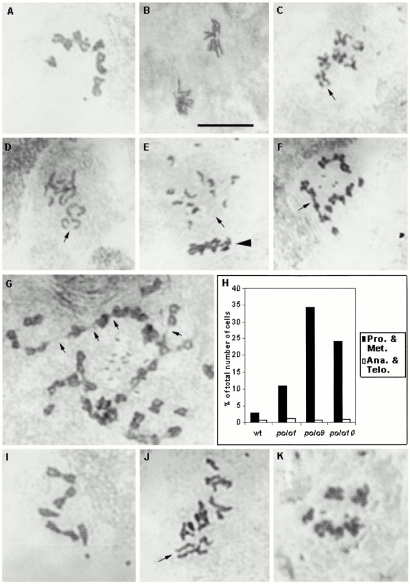
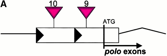
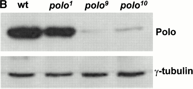
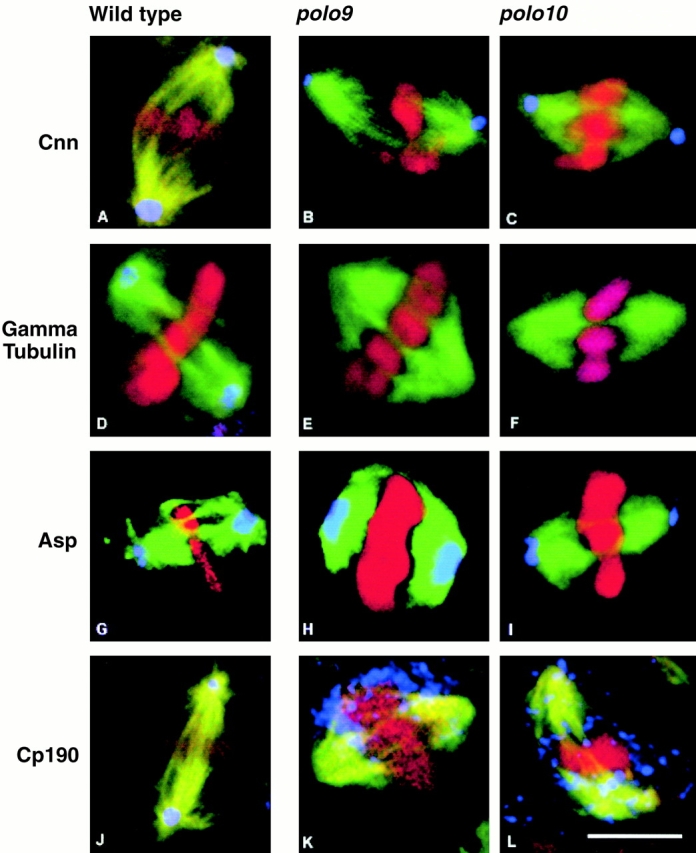
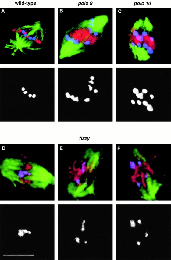
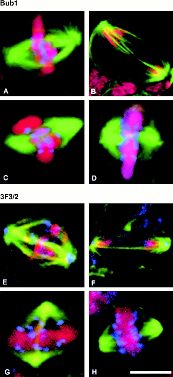
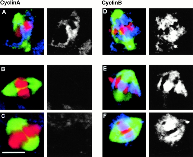
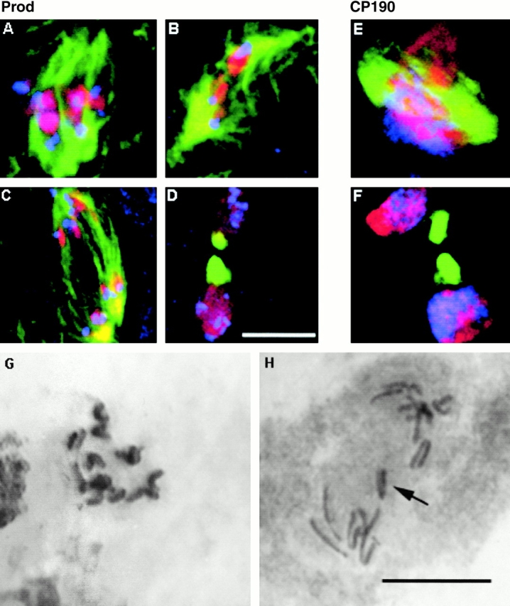
The Drosophila gene polo encodes a conserved protein kinase known to be required to organize spindle poles and for cytokinesis. Here we report two strongly hypomorphic mutations of polo that arrest cells of the larval brain at a point in metaphase when the majority of sister kinetochores have separated by between 20-50% of the total spindle length in intact cells. In contrast, analysis of sister chromatid separation in squashed preparations of cells indicates that some 83% of sisters remain attached. This suggests the separation seen in intact cells requires the tension produced by a functional spindle. The point of arrest corresponds to the spindle integrity checkpoint; Bub1 protein and the 3F3/2 epitope are present on the separated kinetochores and the arrest is suppressed by a bub1 mutation. The mutant mitotic spindles are anastral and have assembled upon centrosomes that are associated with Centrosomin and the abnormal spindle protein (Asp), but neither with gamma-tubulin nor CP190. We discuss roles for Polo kinase in recruiting centrosomal proteins and in regulating progression through the metaphase-anaphase checkpoint.


Figures

Figure 1
Figure 1
Mitotic figures from wild-type, polo 9, and polo 10 brains. (A) A wild-type metaphase figure. (B) A wild-type anaphase figure. (C) Hypercondensed mitotic chromosomes from polo 9 . The arrow points to a pair of separated sister chromatids. (D) polo 10 cell showing separated sisters (arrow) that could be at early anaphase. (E) An anaphase (arrow) and metaphase (arrowhead) figure from polo 10 . (F) A tetraploid polo 10 cell in which several sister chromatids appear separated throughout their length, and yet joined at their telomeres (arrow). (G) A polyploid polo 9 cell in which chromosomes form long chains attached at their telomeres (arrows). (H) Bar graph showing the proportion of cells at prophase and metaphase (Pro. & Met.) or anaphase and telophase (Ana. & Tel.) in wild-type (wt), polo, polo 9, and polo 10 brains. (I) Wild-type cell from a larval brain treated for 4 h with colchicine. (J) polo 10 cell after 30 min colchicine treatment. The arrow marks separated sisters. (K) polo 10 cell after 2 h colchicine treatment.
Figure 2
Figure 2
Sites of P-element insertions in polo 9 and polo 10 and relative expression levels of their gene products. (A) Schematic diagram of insertion sites in which P-elements are indicated by the filled red triangles above the linear map at positions −34 and −176 nucleotides upstream of the initiator ATG codon of the Polo protein. Two major starts for the initiation of transcription are indicated by the horizontal black triangles at positions −130 and −210 nucleotides. Exons are indicated by the open box and the first intron by the kinked line. (B) Western blots to compare the levels of Polo kinase and γ-tubulin in wild-type (wt), polo, polo 9, and polo 10 larval brains (see Materials and Methods).
Figure 2
Figure 2
Sites of P-element insertions in polo 9 and polo 10 and relative expression levels of their gene products. (A) Schematic diagram of insertion sites in which P-elements are indicated by the filled red triangles above the linear map at positions −34 and −176 nucleotides upstream of the initiator ATG codon of the Polo protein. Two major starts for the initiation of transcription are indicated by the horizontal black triangles at positions −130 and −210 nucleotides. Exons are indicated by the open box and the first intron by the kinked line. (B) Western blots to compare the levels of Polo kinase and γ-tubulin in wild-type (wt), polo, polo 9, and polo 10 larval brains (see Materials and Methods).
Figure 3
Figure 3
Distribution of centrosomal antigens in wild-type, polo 9, and polo 10 cells. In all cases, wild-type cells are shown in the left panels, polo 9 cells the middle panels, and polo 10 cells the right panels. Spindle microtubules revealed by immunostaining with the YL1/2 antibody are shown in green, DNA stained with propidium iodide in red, and the centrosomal antigen in blue. (A–C) Centrosomin (Cnn) is revealed using a rabbit antibody from Heuer et al. 1995. (D–F) γ-Tubulin was detected using the mouse monoclonal antibody GTU88 (Sigma-Aldrich). (G–I) The Asp was detected using the rabbit antibody Rb3133 (Saunders et al. 1997). (J–L) CP190 was detected using the rabbit antibody RB188 (Whitfield et al. 1988). Bar, 5 μm.
Figure 4
Figure 4
Localization of Prod in wild-type (A), polo (B and C), and fizzy (D–F) cells. Spindle microtubules stained with the rat monoclonal antibody YL1/2 are stained green. DNA is stained red. Prod (blue) was detected using a rabbit antibody (Torok et al. 1997). Merged images are shown in the top panels with the separated channel for Prod staining below. Bar, 5 μm. 79.3% of 213 clear pairs of sister centromeric regions were scored as having separated in immunostained polo 9 cells. A similar frequency of separation (78.6% of 112 pairs) was observed in polo 10 cells. Centromere separation was not observed by anti-Prod staining in the fizzy x4 mutant.
Figure 5
Figure 5
Association of Bub1 and the 3F3/2 epitope with the mitotic apparatus in wild-type and polo mutant cells. In all panels, spindle microtubules are stained green and DNA stained red. Bub1 (A–F) or 3F3/2 (G–L) staining are shown in blue. (A and B) Wild-type cells at metaphase and anaphase, respectively. (C and D) polo 9 cells showing Bub1 staining with the rabbit antibody Rb666 (gift of C. Sunkel, University of Porto, Porto, Portugal) on the separated kinetochores. (E and F) Wild-type cells at metaphase and anaphase, respectively, stained with the 3F3/2 mouse monoclonal antibody (gift of G. Gorbsky, University of Oklahoma, Oklahoma City, Oklahoma). Note the presence of the 3F3/2 epitope at centrosomes. (G and H) polo 10 cells showing 3F3/2 staining on the separated kinetochores, and absent from the spindle poles. Bar, 5 μm.
Figure 6
Figure 6
Cyclins A and B in wild-type and polo mutant cells. In all panels, spindle microtubules are green, DNA red, and cyclin blue in the merged images. The monochromatic image is of cyclin staining alone using either the antibody Rb270 to detect cyclin A or Rb271 to detect cyclin B (Whitfield et al. 1990). (A) Cyclin A in a wild-type cell at prometaphase. (B and C) Absence of cyclin A staining in polo 9 and polo 10 mutant cells respectively. (D) Cyclin B staining in a wild-type cell at metaphase. (E and F) Cyclin B staining of polo 9 and polo 10 cells, respectively. Bar, 5 μm.
Figure 7
Figure 7
Suppression of the polo 10 mitotic phenotype by bub1. In A–F, microtubules are stained green and DNA stained red. (A–D) polo 10 bub1k6109 cells at metaphase, early anaphase, late anaphase, and telophase, respectively, showing the localization of Prod (blue). (E and F) polo 10 bub1k6109cells at early anaphase and telophase showing the localization of CP190 (blue). Bar, 5 μm. (G and H) Giemsa-stained squashed preparations of polo 10 bub1k6109cells showing, respectively, sister chromatid separation at metaphase and a lagging chromatid (arrow) at anaphase.

References

    1. Abrieu A., Brassac T., Galas S., Fisher D., Labbe J.-C., Doree M. The Polo-like kinase Plx is a component of the MPF amplification loop at the G2/M-phase transition of the cell cycle in Xenopus . J. Cell Sci. 1998;111:1751–1757. - PubMed
    1. Alexandru G., Zachariae W., Schleiffer A., Nasmyth K. Sister chromatid separation and chromosome re-duplication are regulated by different mechanisms in response to spindle damage. EMBO (Eur. Mol. Biol. Organ.) J. 1999;10:2707–2721. - PMC - PubMed
    1. Amon A. The spindle checkpoint. Curr. Opin. Genes Dev. 1999;9:69–75. - PubMed
    1. Avides M., Glover D.M. Abnormal spindle protein Asp, and the integrity of mitotic centrosomal microtubule organizing centres. Science. 1999;283:1733–1735. - PubMed
    1. Barbosa V., Yamamoto R., Carpenter A.T.C., Henderson D.S., Glover D.M. Mutation of a Drosophila gamma tubulin ring complex subunit encoded by discs degenerate-4 differentially disrupts centrosomal protein localization. Genes Dev. 2000;14:3126–3139. - PMC - PubMed

Publication types

MeSH terms