. 2001 May 14;153(4):699-708.
doi: 10.1083/jcb.153.4.699.

Phosphorylation-dependent regulation of ryanodine receptors: a novel role for leucine/isoleucine zippers

Affiliations

Phosphorylation-dependent regulation of ryanodine receptors: a novel role for leucine/isoleucine zippers

S O Marx et al. J Cell Biol. .

Abstract

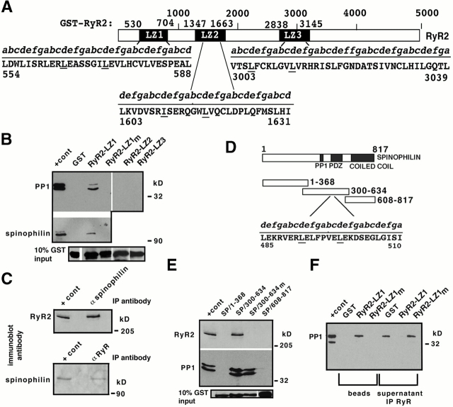
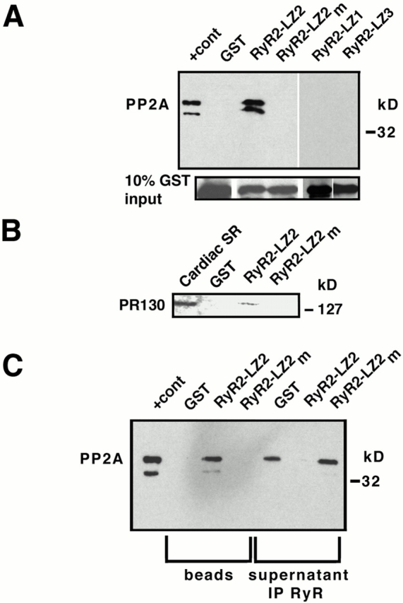
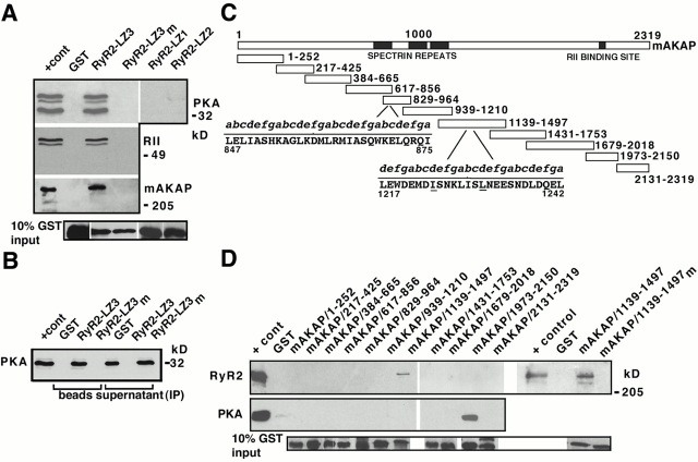
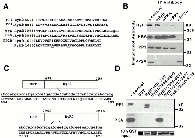
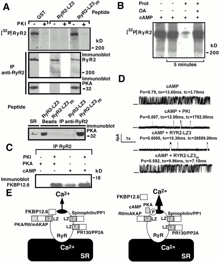
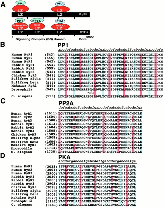
Ryanodine receptors (RyRs), intracellular calcium release channels required for cardiac and skeletal muscle contraction, are macromolecular complexes that include kinases and phosphatases. Phosphorylation/dephosphorylation plays a key role in regulating the function of many ion channels, including RyRs. However, the mechanism by which kinases and phosphatases are targeted to ion channels is not well understood. We have identified a novel mechanism involved in the formation of ion channel macromolecular complexes: kinase and phosphatase targeting proteins binding to ion channels via leucine/isoleucine zipper (LZ) motifs. Activation of kinases and phosphatases bound to RyR2 via LZs regulates phosphorylation of the channel, and disruption of kinase binding via LZ motifs prevents phosphorylation of RyR2. Elucidation of this new role for LZs in ion channel macromolecular complexes now permits: (a) rapid mapping of kinase and phosphatase targeting protein binding sites on ion channels; (b) predicting which kinases and phosphatases are likely to regulate a given ion channel; (c) rapid identification of novel kinase and phosphatase targeting proteins; and (d) tools for dissecting the role of kinases and phosphatases as modulators of ion channel function.


Figures

Figure 1
Figure 1
PP1/spinophilin binds to an LZ motif in RyR2. (A) RyR2–LZ motifs. GST–RyR2-LZ1 (aa 530–704) contains an LZ (aa 555–604); GST-RyR2-LZ2 (aa 1,347–1,663) contains an LZ (aa 1,603–1,631); GST-RyR2-LZ3 (aa 2,838–3,145) contains an LZ (aa 3,003–3,039); underlined residues were substituted with alanines (RyR2–LZXm). (B) Pulldown using cardiac SR (200 μg,) followed by immunoblotting with anti-PP1 or antispinophilin demonstrated specific LZ-dependent binding of PP1 (top panel) and spinophilin (middle panel) to RyR2; “+ cont” represents 5% (spinophilin) or 10% (RyR2) SR input. RyR2–LZ1 but not RyR2–LZ2 or RyR–LZ3 coprecipitated PP1. (C) Coimmunoprecipitation of RyR2 and spinophilin from SR; “+ cont” represents 10% SR input. (D) GST–spinophilin fusion proteins, GST-SP/1–368, GST-SP/300–634, and GST-SP/608–817. GST-SP/300–634 contains the PP1 binding domain (Hsieh-Wilson et al. 1999), a PDZ domain (Allen et al. 1997), and an LZ (aa 485–510). Underlined leucine residues were substituted with alanines (SP/300–634m). (E) Pulldown assays demonstrated a specific interaction between RyR2 and spinophilin; “+ cont” represents either 5% (PP1) or 10% (RyR2) SR input. (F) SR was preincubated with GST alone, RyR2–LZ1, or RyR2–LZ1m before immunoprecipitating supernatants with anti-RyR antibody. Spinophilin/PP1 was competed off RyR2 by GST-RyR2-LZ1, but not by GST alone or RyR2–LZ1m.
Figure 2
Figure 2
PP2A is targeted to RyR2 via an LZ. (A) PP2A specifically bound GST-RyR2-LZ2 (via PR130), but not RyR2–LZ2m in GST-SR pulldown assays. RyR2–LZ2 but not RyR2–LZ1 or RyR–LZ3 coprecipitated PP2A. “+ cont” represents 5% SR input. (B) PR130 specifically bound GST-RyR2-LZ2, but not RyR2–LZ2m in GST-SR pulldown assays. “Cardiac SR” represents 5% SR input. (C) SR was incubated with GST alone, GST-RyR2-LZ2, or GST-RyR2-LZ2m before immunoprecipitating the supernatant with anti-RyR antibody and immunoblotting with anti-PP2A. PP2A binding to RyR2 was competitively inhibited by incubation with GST-RyR2-LZ2.
Figure 3
Figure 3
PKA/RII/mAKAP bind to RyR2 via LZ motifs. (A) GST-SR pulldown demonstrated a specific interaction between mAKAP/RII/PKA and RyR2–LZ3, but not GST or GST-RyR2-LZ3m; immunoblots, anti-PKA (first panel), anti-RII (second panel), and anti-mAKAP (third panel). “+ cont” represents 5% (RII and PKA) or 10% (mAKAP) SR input. (B) SR was incubated with GST, GST-RyR2-LZ3, or GST-RyR2-LZ3m before immunoprecipitation of supernatants with anti-RyR antibody followed by immunoblotting with anti-PKA. RyR2–LZ3 but not RyR2–LZ1 or RyR–LZ2 coprecipitated PKA. Lanes labeled “beads” contain GST-pulldown samples, and lanes labeled “supernatant” are immunoprecipitations. PKA binding to RyR2 via mAKAP was competitively inhibited by incubation with GST-RyR2-LZ3 but not GST or GST-RyR2-LZ3m. “+ cont” represents 5% SR input. (C) mAKAP and GST-mAKAP fusion proteins. GST-mAKAP/1,973–2,150 contains the RII binding domain, GST-mAKAP/829–964 and GST-mAKAP/1,139–1,497 contain LZ motifs. Alanines were substituted for underlined isoleucine/leucines (GST-mAKAP/1,139–1,497m). (D) GST-SR pulldown assays followed by immunoblotting with anti-RyR (top) or anti-PKA (middle). “+ cont” represents 10% (RyR2, top) and 5% (PKA, middle) SR input.
Figure 5
Figure 5
Components of the RyR1 macromolecular signaling complex bind via LZ motifs. (A) Comparison of aa sequences required for targeting of PP1, PKA, and PP2A to RyR2 and PP1 and PKA to RyR1. The second and third “d” position of RyR2–PP2A targeting motif is not conserved in RyR1 (underlined residues), implying that PP2A is not targeted to RyR1 through this motif. Numbers in parentheses correspond to the first residue in each sequence. (B) RyR1 macromolecular signaling complex contains PKA and PP1, but not PP2A. Components of the RyR1 complex, PKA and PP1, were coimmunoprecipitated from purified skeletal SR (200 μg; Marx et al. 1998). Positive (+) control represents 5% (PKA, PP1, and PP2A) and 10% (RyR) of SR input. Data shown are representative of more than three similar experiments. (C) Schematic diagram of PP1 and PKA binding domain on RyR1. The GST fusion protein corresponding to aa 497–759 of RyR1 contains a leucine/valine heptad repeat between aa 554–603. The GST fusion protein corresponding to aa 2,968–3,216 of RyR1 contains a leucine/valine heptad repeat between aa 3,039–3,075. The motif is comprised of a periodic repeat (labeled “abcdefg”) of a leucine at every seventh or “d” position with a “skip” labeled “ff” between repeats 3 and 4. (D) GST pulldown assays demonstrated a specific interaction between RyR1 and PP1 and RyR1 and PKA through putative LZ targeting motifs. GST fusion proteins were incubated with rabbit skeletal SR membranes (200 μg). Bound proteins were analyzed with SDS-PAGE. The positive (+) control represents 5% of SR input. GST fusion protein, GST-RyR1/497–759 demonstrated specific interaction with PP1 (top panel). GST fusion protein, GST-RyR1/2,968–3,216 demonstrated specific interaction with PKA (middle panel). Separate immunoblots containing 10% of the GST fusion protein input were probed with anti-GST antibody to demonstrate that equivalent amounts of GST fusion proteins were used in pulldown assays (bottom panel). As GST fusion proteins are different molecular weights, bands were cut from immunoblot and realigned for purposes of comparison.
Figure 4
Figure 4
Kinase and phosphatases bound to RyR2 via LZ motifs regulate channel function. (A) Top panel: immunoprecipitated RyR2 was phosphorylated via activation of bound kinase by addition of cAMP (10 μM) and [γ-32P]ATP with or without PKI5–24 (500 nM). RyR2 phosphorylation by bound PKA was inhibited by preincubation with GST-RyR2-LZ3, but not by incubation with either GST alone or GST-RyR2-LZ3m (top panel). Equivalent amounts of RyR2 protein were present in each sample (second panel). Incubation with GST-RyR2-LZ3 but not with GST-RyR2-LZ3m disrupted binding of PKA to RyR2 (via mAKAP) as shown by coimmunoprecipitation (third panel). After incubation with GST-RyR2-LZ3, the PKA previously bound to RyR2 was now bound to GST-RyR-LZ3 beads and none was detected after coimmunoprecipitation with RyR2 (bottom panel). After incubation with RyR2–LZ3m, PKA coimmunoprecipitated with RyR2 and none was detected, by immunoblot, bound to the GST-RyR-LZ3m beads (bottom panel). “SR” denotes a sample of cardiac SR used as a positive control for the anti-PKA antibody. (B) RyR2-bound phosphatases dephosphorylate RyR2. Immunoprecipitated RyR2 was phosphorylated with cAMP for 15 min, and further phosphorylation was inhibited by PKI. RyR2 was dephosphorylated for 5 min by activation of bound phosphatases with protamine (Prot; 1 mg/ml), which was inhibited by okadaic acid (OA; 5 nM). (C) cAMP-induced activation of RyR2-bound PKA caused dissociation of FKBP12.6. (D) Pelleted membranes were introduced into planar lipid bilayers and RyR2 single channel properties determined. cAMP-induced phosphorylation of RyR2 by bound PKA increased P o. This was inhibited by the PKA inhibitor PKI. Preincubation with GST-RyR2-LZ3 (RYR2–LZ3) but not with GST-RyR2-LZ3m (RYR2–LZ3m) disrupted binding of PKA to RyR2 (see A) and inhibited the cAMP-dependent increase in channel P o. (E) Model of RyR2 macromolecular signaling complex, one subunit of the tetrameric RyR channel is shown. Targeting of kinases and phosphatases to RyR2 requires LZ interactions between RyR2 and targeting proteins. cAMP activates bound PKA, phosphorylating RyR2 and increasing calcium release.
Figure 6
Figure 6
Conservation of LZ motifs in RyRs. (A) Location of the LZ motifs on RyR1 and RyR2 that bind to LZ motifs in adaptor/targeting proteins for PKA, PP1, and PP2A. The numbers below the RyR2 refer to aa residues. RyR sequences corresponding to the LZ motifs that bind (B) PP1, (C) PP2A, and (D) PKA were aligned. Numbers in parentheses correspond to the first residue in each sequence. Blue shaded residues indicate conserved “a” and red shaded residues indicate conserved “d” positions of the leucine/isoleucine heptad repeats. There are three forms of RyR identified to date, RyR1 (Marks et al. 1989; Takeshima et al. 1989; Zorzato et al. 1990), RyR2 (Nakai et al. 1990; Otsu et al. 1990), and RyR3 (Hakamata et al. 1992). The frog alpha corresponds to RyR1 and beta to RyR3 (Oyamada et al. 1994). The fish RyR (blue marlin, Makaira nigricans) corresponds to RyR1 (Franck et al. 1998). The Drosophila RyR is ∼45% homologous to mammalian RyRs (Takeshima et al. 1994; Xu et al. 2000), and C. elegans RyR corresponds to RyR1 (Maryon et al. 1996). In B, the (*) indicates two residues inserted in the Drosophila sequence that displace the alignment of the LZ motif. The proline in the middle of the PP1 LZ motif would introduce a bend in the helix (Hendrickson and Love 1971) which is followed by a phase shift “skip” (eight residues instead of seven between the leucine/isoleucine in the repeat). The phase shift could bring the repeats back into register following the bend in the helix to permit alignment with the corresponding heptad repeats in the LZ motif in spinophilin.

References

    1. Alber T., Bell J.A., Sun D.P., Nicholson H., Wozniak J.A., Cook S., Matthews B.W. Replacements of Pro86 in phage T4 lysozyme extend an alpha-helix but do not alter protein stability. Science. 1988;239:631–635. - PubMed
    1. Allen P., Ouimet C., Greengard P. Spinophilin, a novel protein phosphatase 1 binding protein localized to dendritic spines. Proc. Natl. Acad. Sci. USA. 1997;94:9956–9961. - PMC - PubMed
    1. Angelo R., Rubin C.S. Molecular characterization of an anchor protein (AKAPCE) that binds the RI subunit (RCE) of type I protein kinase A from Caenorhabditis elegans . J. Biol. Chem. 1998;273:14633–14643. - PubMed
    1. Barlow D.J., Thornton J.M. Helix geometry in proteins. J. Mol. Biol. 1988;201:601–619. - PubMed
    1. Brillantes A.B., Ondrias K., Scott A., Kobrinsky E., Ondriasova E., Moschella M.C., Jayaraman T., Landers M., Ehrlich B.E., Marks A.R. Stabilization of calcium release channel (ryanodine receptor) function by FK506-binding protein. Cell. 1994;77:513–523. - PubMed

Publication types

MeSH terms