. 2001 May 14;153(4):725-34.
doi: 10.1083/jcb.153.4.725.

Intramitochondrial localization of universal minicircle sequence-binding protein, a trypanosomatid protein that binds kinetoplast minicircle replication origins

Affiliations

Intramitochondrial localization of universal minicircle sequence-binding protein, a trypanosomatid protein that binds kinetoplast minicircle replication origins

K Abu-Elneel et al. J Cell Biol. .

Abstract

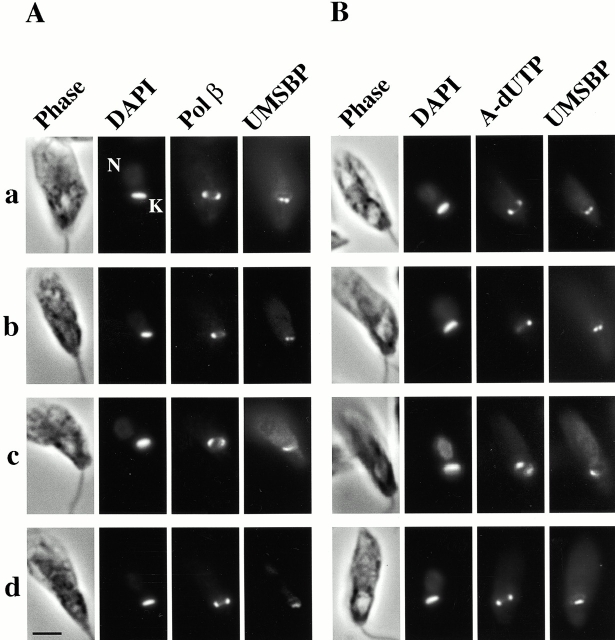
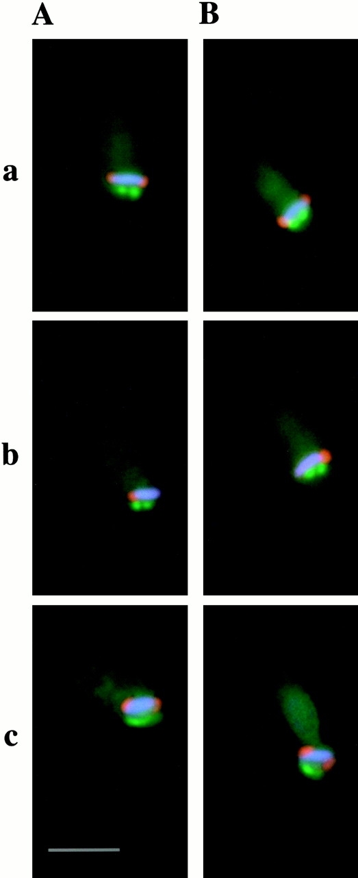
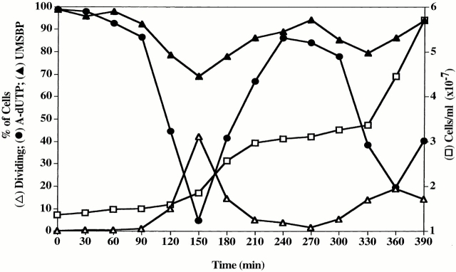
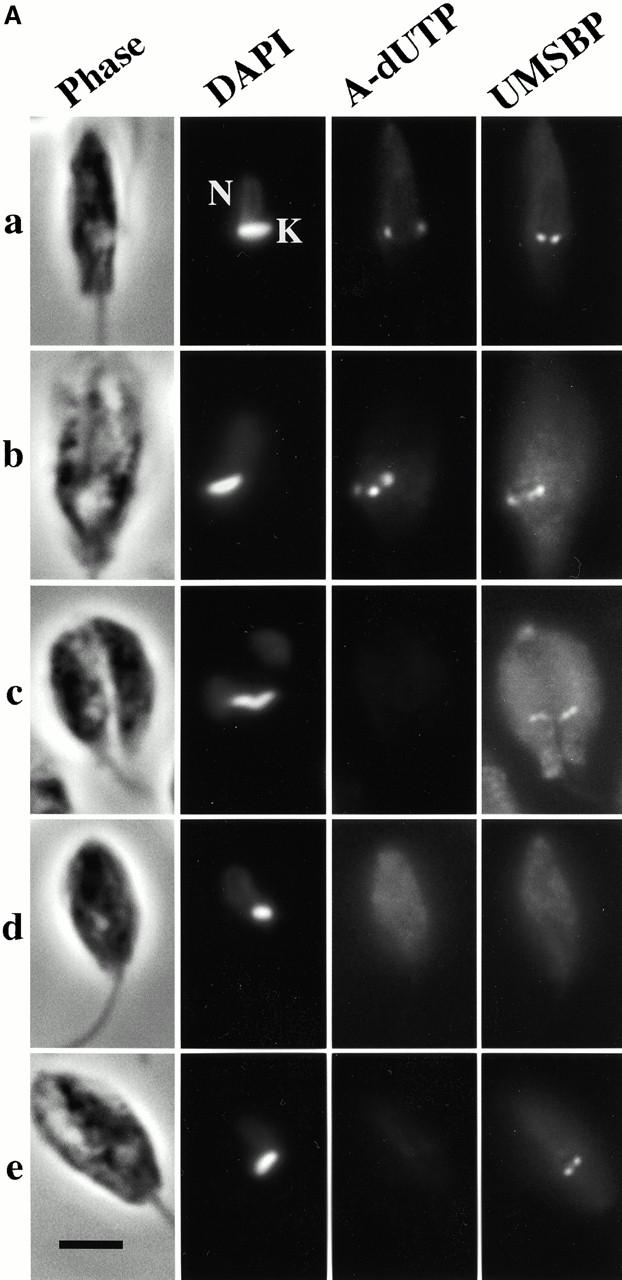
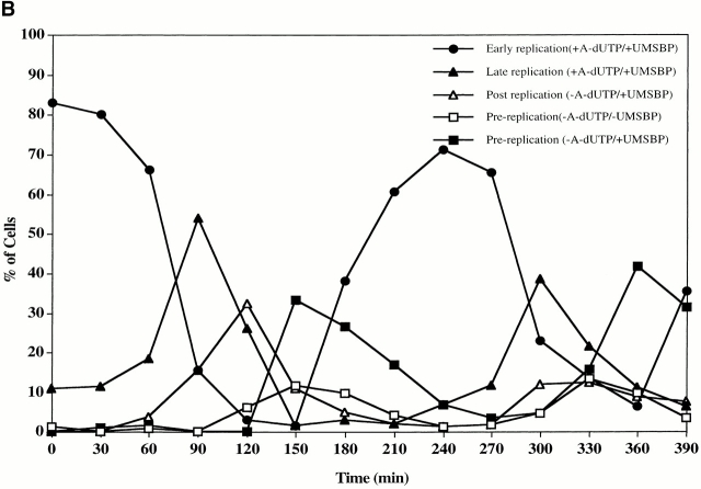
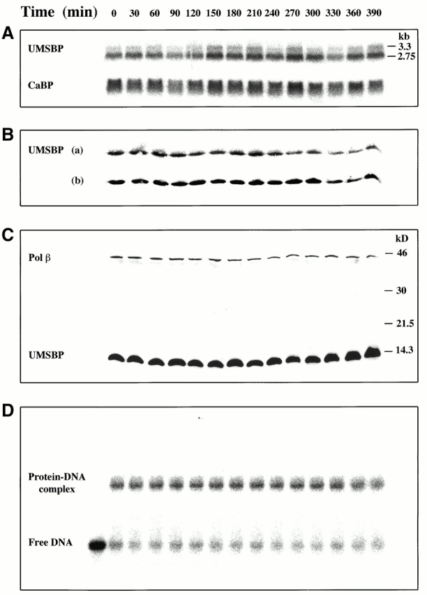
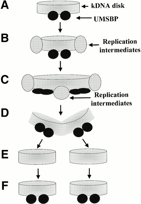
Kinetoplast DNA (kDNA), the mitochondrial DNA of the trypanosomatid Crithidia fasciculata, is a unique structure containing 5,000 DNA minicircles topologically linked into a massive network. In vivo, the network is condensed into a disk-shaped structure. Replication of minicircles initiates at unique origins that are bound by universal minicircle sequence (UMS)-binding protein (UMSBP), a sequence-specific DNA-binding protein. This protein, encoded by a nuclear gene, localizes within the cell's single mitochondrion. Using immunofluorescence, we found that UMSBP localizes exclusively to two neighboring sites adjacent to the face of the kDNA disk nearest the cell's flagellum. This site is distinct from the two antipodal positions at the perimeter of the disk that is occupied by DNA polymerase beta, topoisomerase II, and a structure-specific endonuclease. Although we found constant steady-state levels of UMSBP mRNA and protein and a constant rate of UMSBP synthesis throughout the cell cycle, immunofluorescence indicated that UMSBP localization within the kinetoplast is not static. The intramitochondrial localization of UMSBP and other kDNA replication enzymes significantly clarifies our understanding of the process of kDNA replication.


Figures

Figure 1
Figure 1
Immunolocalization of UMSBP in an asynchronous C. fasciculata cell culture. Phase and fluorescence microscopy are described in Materials and Methods. With DAPI staining, the kDNA disk displays bright fluorescence, and the nucleus fluoresces weakly. (A) Immunofluorescence of pol β and UMSBP. For pol β, the secondary antibody was goat anti–rabbit conjugated to Texas red, and for UMSBP it was rat anti–mouse conjugated to FITC. (B) Immunofluorescence of UMSBP in cells labeled in situ with Alexa-dUTP (using TdT as described in Materials and Methods). a, b, and d present edgewise views of the kDNA disk, whereas c shows a disk that is partially tipped over. K, kDNA; N, nucleus; A-dUTP, Alexa-dUTP. Bar, 3 μm.
Figure 2
Figure 2
Localization of UMSBP, pol β, and minicircle replication intermediates. Phase and fluorescence microscopy were conducted as described in Materials and Methods. (A) An overlay presentation of DAPI staining (blue), pol β (red), and UMSBP (green) immunostaining of the cell images presented in Fig. 1 A, a–c. (B) An overlay presentation of DAPI staining (blue), Alexa-dUTP fluorescence (red), and UMSBP (green) of the cell images presented in Fig. 1 B, a–c. Bar, 3 μm.
Figure 3
Figure 3
UMSBP in synchronized cell cultures. Cell cultures were synchronized as described previously (Pasion et al. 1994). Cells (107 cells/ml) were incubated for 6 h at 26°C in the presence of 200 μg/ml hydroxyurea, centrifuged (6,000 g, 15 min), resuspended in fresh culture medium, and grown for an additional 6.5 h. Samples were withdrawn at the indicated times and counted (open squares). The percent of cells undergoing division containing two nuclei was determined by fluorescence and phase microscopy (open triangles), and the percent of cells displaying UMSBP (filled triangles) and Alexa-dUTP (A-dUTP) (filled circles) fluorescence was determined by fluorescence microscopy.
Figure 4
Figure 4
Variations in the intraorganelle localization of UMSBP during the cell cycle. Cell cultures were as described in the legend to Fig. 3. Phase and fluorescence microscopy were conducted as described in Materials and Methods. Phase microscopy, DAPI fluorescence, and immunofluorescence of UMSBP in cells labeled in situ with fluorescent Alexa-dUTP are presented. (A) Typical examples of cells sampled at various times after release from hydroxyurea arrest. (a) Cell at an early stage of kDNA replication (at 30 min and then at the subsequent cell cycle at 240 min after the release from hydroxyurea arrest); (b) cell at a late stage of kDNA replication (90 and then 300 min); (c) cell at a postreplication stage (120 and then 300–330 min); (d and e) cells in a prereplication stage (150–180 and then 330–360 min). (B) Quantification of cell images throughout two consecutive cell cycles. Cells displaying both Alexa-dUTP and UMSBP fluorescence at an early stage of kDNA replication, like that in A (a, filled circles), or a late stage, like that in A (b, filled triangles). Cells displaying UMSBP fluorescence but little or no Alexa-dUTP fluorescence at a post-kDNA replication stage, like that in A (c, open triangles). Cells at a stage before undergoing kDNA replication (no Alexa-dUTP labeling), either with UMSBP, like that in A (e, filled squares), or without UMSBP, like that in A (d, open squares). K, kDNA; N, nucleus; A-dUTP, Alexa-dUTP. Bar, 3 μm.
Figure 4
Figure 4
Variations in the intraorganelle localization of UMSBP during the cell cycle. Cell cultures were as described in the legend to Fig. 3. Phase and fluorescence microscopy were conducted as described in Materials and Methods. Phase microscopy, DAPI fluorescence, and immunofluorescence of UMSBP in cells labeled in situ with fluorescent Alexa-dUTP are presented. (A) Typical examples of cells sampled at various times after release from hydroxyurea arrest. (a) Cell at an early stage of kDNA replication (at 30 min and then at the subsequent cell cycle at 240 min after the release from hydroxyurea arrest); (b) cell at a late stage of kDNA replication (90 and then 300 min); (c) cell at a postreplication stage (120 and then 300–330 min); (d and e) cells in a prereplication stage (150–180 and then 330–360 min). (B) Quantification of cell images throughout two consecutive cell cycles. Cells displaying both Alexa-dUTP and UMSBP fluorescence at an early stage of kDNA replication, like that in A (a, filled circles), or a late stage, like that in A (b, filled triangles). Cells displaying UMSBP fluorescence but little or no Alexa-dUTP fluorescence at a post-kDNA replication stage, like that in A (c, open triangles). Cells at a stage before undergoing kDNA replication (no Alexa-dUTP labeling), either with UMSBP, like that in A (e, filled squares), or without UMSBP, like that in A (d, open squares). K, kDNA; N, nucleus; A-dUTP, Alexa-dUTP. Bar, 3 μm.
Figure 5
Figure 5
Expression of UMSBP throughout the cell cycle. C. fasciculata cell cultures were synchronized as described in the legend to Fig. 3. In A, the steady-state levels of UMSBP mRNA in synchronized cell cultures were determined by Northern blot hybridization analyses of total cell RNA (15 μg/lane) isolated at 30 min intervals after the release from hydroxyurea arrest using UMSBP and CaBP ORFs probes as described in Materials and Methods. In B, the rate of UMSBP synthesis during the cell cycle was followed in synchronized cell cultures, monitoring the incorporation of [35S]methionine/cysteine during short (15 min) pulse labeling at the indicated time intervals after the release of hydroxyurea arrest. Metabolic labeling of C. fasciculata cultures was conducted as described in Materials and Methods. Sampled cells were lysed, and the cleared cell lysates were immunoprecipitated with anti-UMSBP antibody and protein A–Sepharose and analyzed by electrophoresis in 16.5% Tris-tricine SDS-polyacrylamide gels as described in Materials and Methods. Indicated are 35S-labeled immunoprecipitates of UMSBP quantified by phosphorImager (a) relative to their respective Western blots developed by ECL and quantified by densitometry (b). In C, the abundance of UMSBP in synchronized C. fasciculata cell cultures was determined by Western blot analyses as described in Materials and Methods. Cells withdrawn at the indicated time intervals after the release from hydroxyurea arrest were lysed, and cleared cell lysates were electrophoresed (20 μg protein/lane), blotted onto membranes, and probed with anti-UMSBP and anti-pol β antibodies. Detection of immune complexes by ECL was described in Materials and Methods. In D, UMSBP activity in synchronized cell cultures was conducted by the electrophoretic mobility shift analysis as described in Materials and Methods. Lysates (fraction I; Tzfati et al. 1995) of cell aliquots withdrawn at the indicated time intervals were used (10 ng protein/assay), and 12 fmol of the 32P-labeled UMS DNA ligand were used. Indicated are UMSBP-UMS DNA complexes and free UMS DNA.
Figure 6
Figure 6
Schematic description of UMSBP localization throughout the cell cycle. The scheme is based upon fluorescence microscopy of UMSBP immunolocalization, DAPI staining of kDNA networks, and incorporation of fluorescently labeled dUTP into gapped kDNA replication intermediates in synchronized cell cultures. See text for details. Note that in these diagrams, the basal body of the flagellum is positioned just below the kDNA disk.

References

    1. Abeliovich H., Tzfati Y., Shlomai J. A trypanosomal CCHC-type zinc finger protein which binds the conserved universal sequence of kinetoplast DNA minicirclesisolation and analysis of the complete cDNA from Crithidia fasciculata . Mol. Cell. Biol. 1993;13:7766–7773. - PMC - PubMed
    1. Abu-Elneel K., Kapeller I., Shlomai J. Universal minicircle sequence-binding protein, a sequence-specific DNA-binding protein that recognizes the two replication origins of the kinetoplast DNA minicircle. J. Biol. Chem. 1999;274:13419–13426. - PubMed
    1. Avrahami D., Tzfati Y., Shlomai J. A single-stranded DNA binding protein binds the origin of replication of the duplex kinetoplast DNA. Proc. Natl. Acad. Sci. USA. 1995;92:10511–10515. - PMC - PubMed
    1. Carpenter L.R., Englund P.T. Kinetoplast maxicircle DNA replication in Crithidia fasciculata and Trypanosoma brucei . Mol. Cell. Biol. 1995;15:6794–6803. - PMC - PubMed
    1. Cosgrove W.B., Skeen M.J. The cell cycle in Crithidia fasciculata. Temporal relationships between synthesis of deoxyribonucleic acid in the nucleus and in the kinetoplast. J. Protozool. 1970;17:172–177. - PubMed

Publication types

MeSH terms