. 2001 May 14;153(4):763-72.
doi: 10.1083/jcb.153.4.763.

Alterations at the intercalated disk associated with the absence of muscle LIM protein

Affiliations

Alterations at the intercalated disk associated with the absence of muscle LIM protein

E Ehler et al. J Cell Biol. .

Abstract

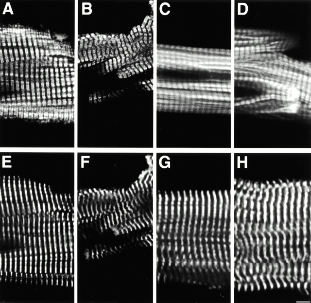
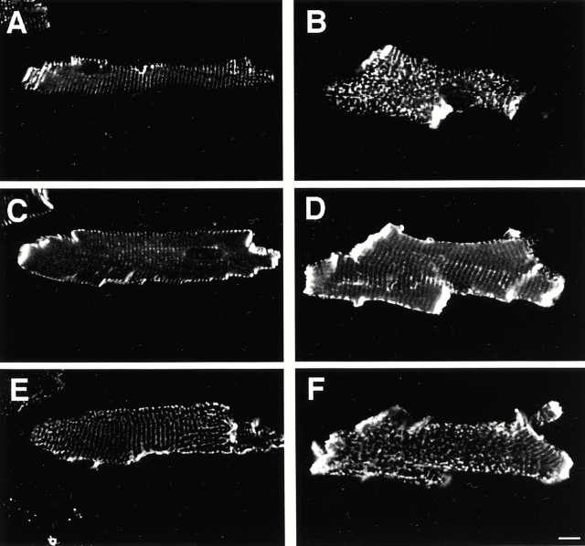
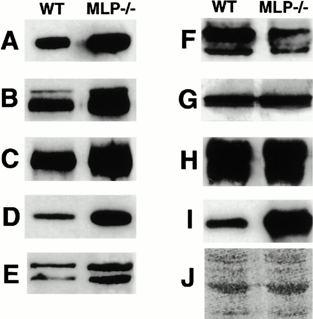
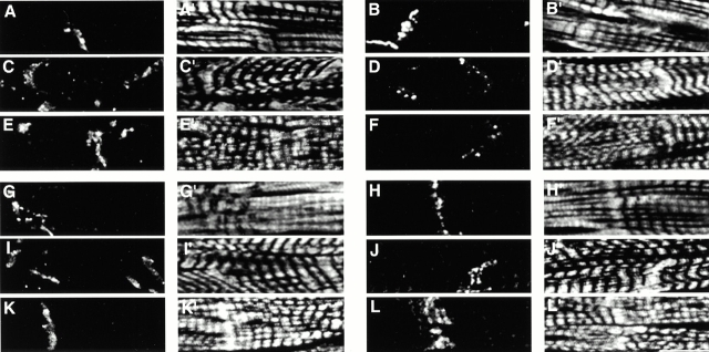
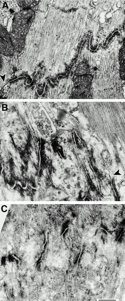
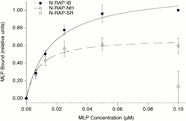
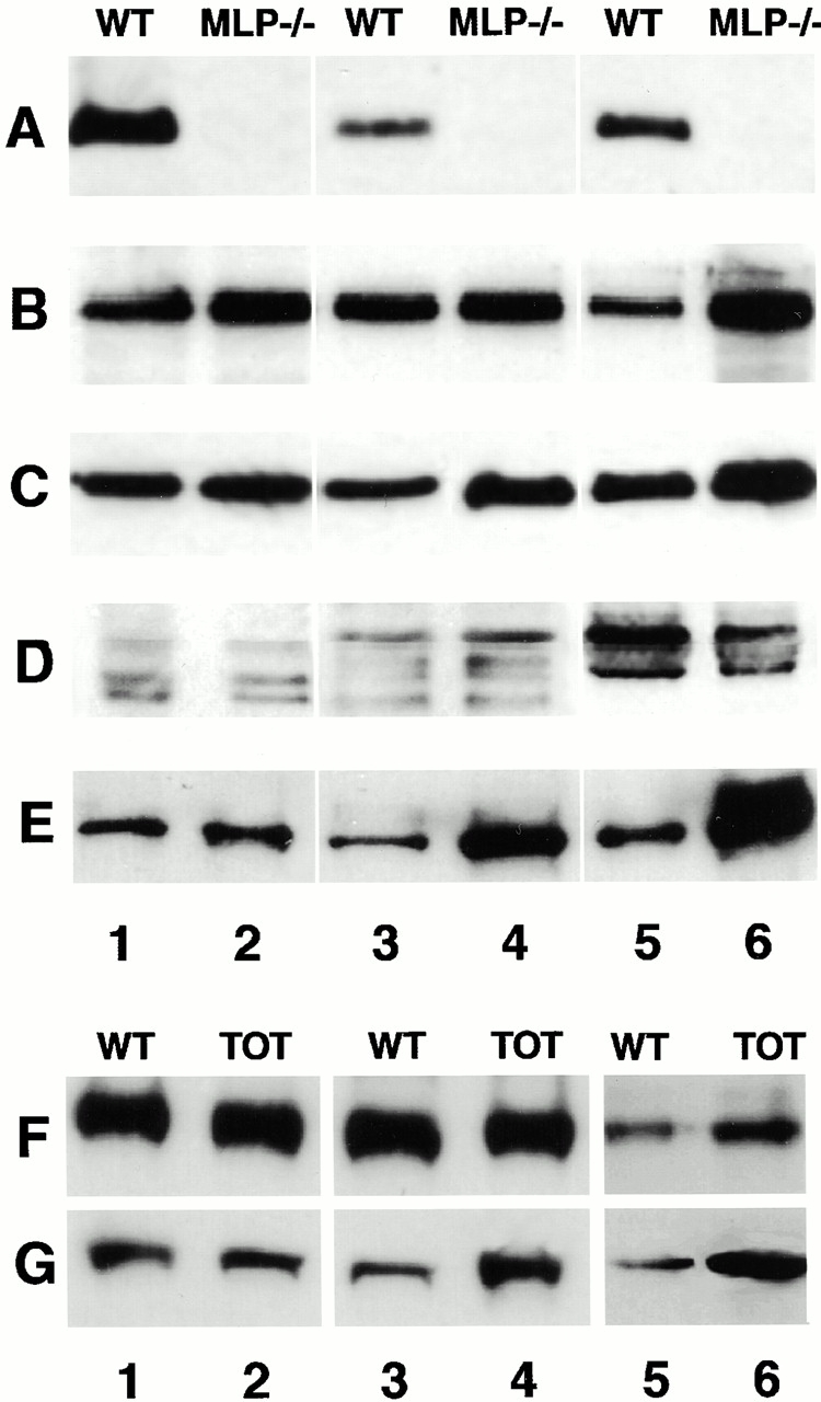
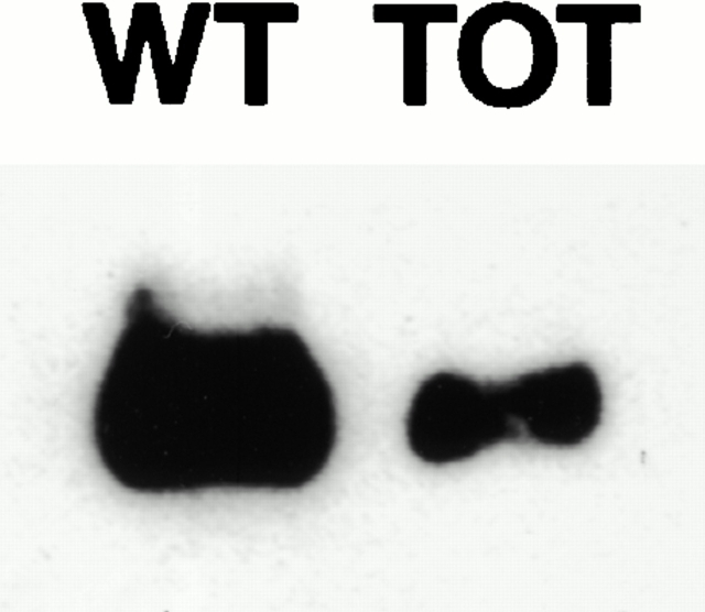
In this study, we investigated cardiomyocyte cytoarchitecture in a mouse model for dilated cardiomyopathy (DCM), the muscle LIM protein (MLP) knockout mouse and substantiated several observations in a second DCM model, the tropomodulin-overexpressing transgenic (TOT) mouse. Freshly isolated cardiomyocytes from both strains are characterized by a more irregular shape compared with wild-type cells. Alterations are observed at the intercalated disks, the specialized areas of mechanical coupling between cardiomyocytes, whereas the subcellular organization of contractile proteins in the sarcomeres of MLP knockout mice appears unchanged. Distinct parts of the intercalated disks are affected differently. Components from the adherens junctions are upregulated, desmosomal proteins are unchanged, and gap junction proteins are downregulated. In addition, the expression of N-RAP, a LIM domain- containing protein located at the intercalated disks, is upregulated in MLP knockout as well as in TOT mice. Detailed analysis of intercalated disk composition during postnatal development reveals that an upregulation of N-RAP expression might serve as an early marker for the development of DCM. Altered expression levels of cytoskeletal proteins (either the lack of MLP or an increased expression of tropomodulin) apparently lead to impaired function of the myofibrillar apparatus and to physiological stress that ultimately results in DCM and is accompanied by an altered appearance and composition of the intercalated disks.


Figures

Figure 1
Figure 1
Sarcomeric organization is normal in freshly isolated cardiomyocytes from MLP−/− mice. Confocal microscope images of freshly isolated cardiomyocytes from wild-type (A, C, E, and G) and MLP−/− mice (B, D, F, and H) stained for components of the A-band (MyBP-C, A and B), the I-band (F-actin visualized by rhodamine–phalloidin in C and D), the Z-disk (sarcomeric α-actinin; E and F), and the M-band (myomesin; G and H). No alterations in the cross-striations were observed; however, the individual myofibrils do not run as strictly parallel in the MLP−/− cardiomyocytes as they do in wild type. Bar, 5 μm.
Figure 2
Figure 2
The organization of the costameric protein vinculin is disturbed in MLP−/− cardiomyocytes. Single confocal sections of freshly isolated cardiomyocytes from wild-type (A, C, and E) and MLP−/− mice (B, D, and F) stained with antibodies to vinculin. Optical sections were taken from the bottom (A and B), through the middle of the cell (C and D), and from the top (E and F). MLP−/− cardiomyocytes display a more irregular overall shape and, instead of a striated arrangement, they show a patchy distribution of vinculin at their surface.
Figure 3
Figure 3
Expression levels of intercalated disk proteins are changed in adult MLP−/− hearts. Immunoblots on whole heart muscle samples of wild-type (WT) or MLP−/− mice with antibodies against cadherin (A), β-catenin (B), plakoglobin (C), α-catenin (D), vinculin (E), connexin-43 (F), desmoplakin (G), desmoglein (H), and N-RAP (I). J shows a part of a Ponceau-stained filter. Adherens junction–associated proteins appear upregulated (A–E, and H); the gap junction component connexin-43 seems downregulated (F), and the expression levels of the desmosomal markers desmoplakin and desmoglein are unaffected (G). Representative blots with extracts from one animal per lane are shown.
Figure 4
Figure 4
Changes of intercalated disk morphology as seen by immunofluorescence. Semithin cryosections of wild type (A, A′, B, B′, G, G′, H, and H′); MLP−/− (C, C′, D, D′, I, I′, J, and J′) and TOT mice (E, E′, F, F′, K, K′, L, and L′) were stained with antibodies against β-catenin (A, C, and E), connexin-43 (B, D, and F), desmoplakin (G, I, and K), and N-RAP (H, J, and L), together with Cy5-phalloidin, to visualize F-actin (A′–L′). Although an increased signal can be observed in the case of adherens junction proteins like β-catenin in DCM hearts, the signal for the gap junction protein connexin-43 appears reduced, and that for desmoplakin as a desmosomal marker is unchanged. In wild-type hearts, N-RAP stains only a single band at the intercalated disk, whereas in DCM hearts, a duplication of this band can be observed. Bar, 10 μm.
Figure 5
Figure 5
Electron microscopy of the altered intercalated disk morphology in MLP−/− hearts. Negatively stained resin sections of wild-type (A), MLP−/− (B), and TOT hearts (C). Compared with wild-type, the intercalated disks of MLP−/−, as well as of TOT mice, are much more convoluted and only rarely show gap junctions (arrowheads). Bar, 500 nm.
Figure 7
Figure 7
Interaction between MLP and N-RAP in a solid phase binding assay. Significant binding of recombinantly expressed full-length MLP can be observed to N-RAP-IB (the region between the LIM domain and the nebulin-like repeats) as well as to N-RAP-NH (N-RAP-IB plus the NH2-terminal LIM domain). The apparent dissociation constants are 17 ± 3 and 8 ± 2 nM, respectively. No significant binding is observed to the N-RAP superrepeats (N-RAP-SR).
Figure 6
Figure 6
Changes in expression levels of intercalated disk proteins during postnatal development. Immunoblots of whole heart muscle samples of wild-type (WT) and MLP−/− mice as well as of TOT of different developmental stages (newborn, lanes 1 and 2; 21-d-old, lanes 3 and 4; adult, lanes 5 and 6) with antibodies against MLP (A), cadherin (B and F), plakoglobin (C), connexin-43 (D), and N-RAP (E and G). Although the differences in the expression levels of most intercalated disk proteins only become apparent by the adult stage (cadherin, plakoglobin, and connexin-43), N-RAP expression is already elevated in samples from newborn MLP−/− mice as well as from juvenile TOT mice compared with wild type. Representative blots with extracts from one animal per lane are shown.
Figure 8
Figure 8
MLP expression is downregulated in hearts from adult TOT mice. Immunoblots on ventricular samples from adult TOT mice (right) and their respective wild-type strain (left) with antibodies specific for MLP reveal a decrease in MLP expression at this developmental stage. Representative blots with extracts from one animal per lane are shown.

References

    1. Angst B.D., Khan L.U., Severs N.J., Whitely K., Rothery S., Thompson R.P., Magee A.I., Gourdie R.G. Dissociated spatial patterning of gap junctions and cell adhesion junctions during postnatal differentiation of ventricular myocardium. Circ. Res. 1997;80:88–94. - PubMed
    1. Arber S., Caroni P. Specificity of single LIM motifs in targeting and LIM/LIM interactions in situ. Genes Dev. 1996;10:289–300. - PubMed
    1. Arber S., Halder G., Caroni P. Muscle LIM protein, a novel essential regulator of myogenesis, promotes myogenic differentiation. Cell. 1994;79:221–231. - PubMed
    1. Arber S., Hunter J.J., Ross J.J., Hongo M., Sansig G., Borg J., Perriard J.-C., Chien K.R., Caroni P. MLP-deficient mice exhibit a disruption of cardiac cytoarchitectural organization, dilated cardiomyopathy, and heart failure. Cell. 1997;88:393–403. - PubMed
    1. Bähler M., Moser H., Eppenberger H.M., Wallimann T. Heart c-protein is transiently expressed during skeletal muscle development in the embryo, but persists in cultured myogenic cells. Dev. Biol. 1985;112:345–352. - PubMed

Publication types

MeSH terms