. 2001 May 14;153(4):773-84.
doi: 10.1083/jcb.153.4.773.

Synergism of Xist RNA, DNA methylation, and histone hypoacetylation in maintaining X chromosome inactivation

Affiliations

Synergism of Xist RNA, DNA methylation, and histone hypoacetylation in maintaining X chromosome inactivation

G Csankovszki et al. J Cell Biol. .

Abstract

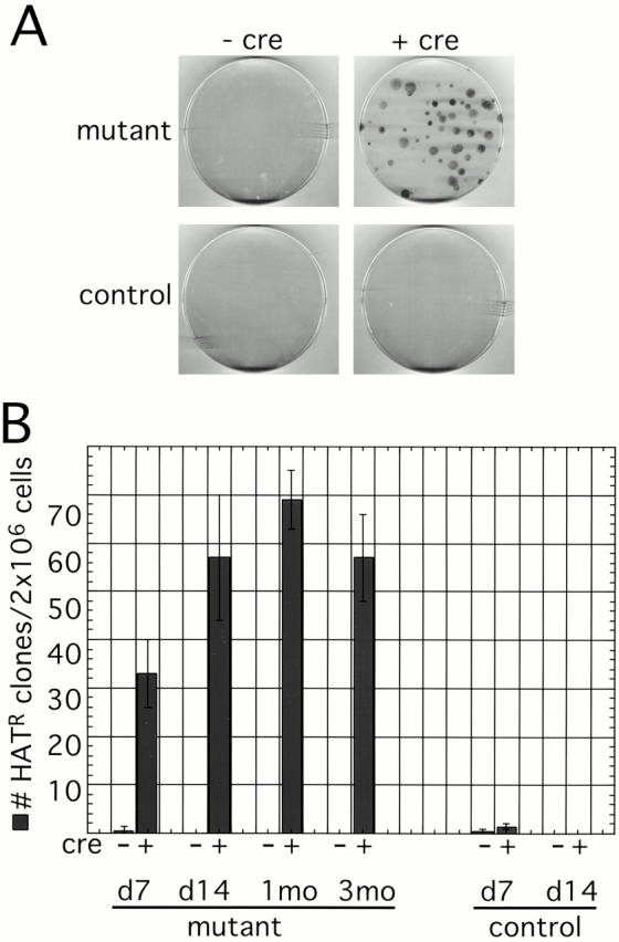
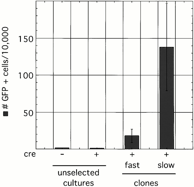
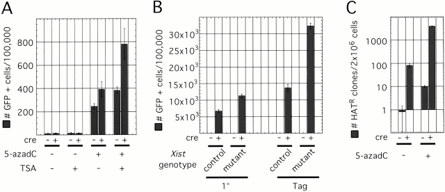
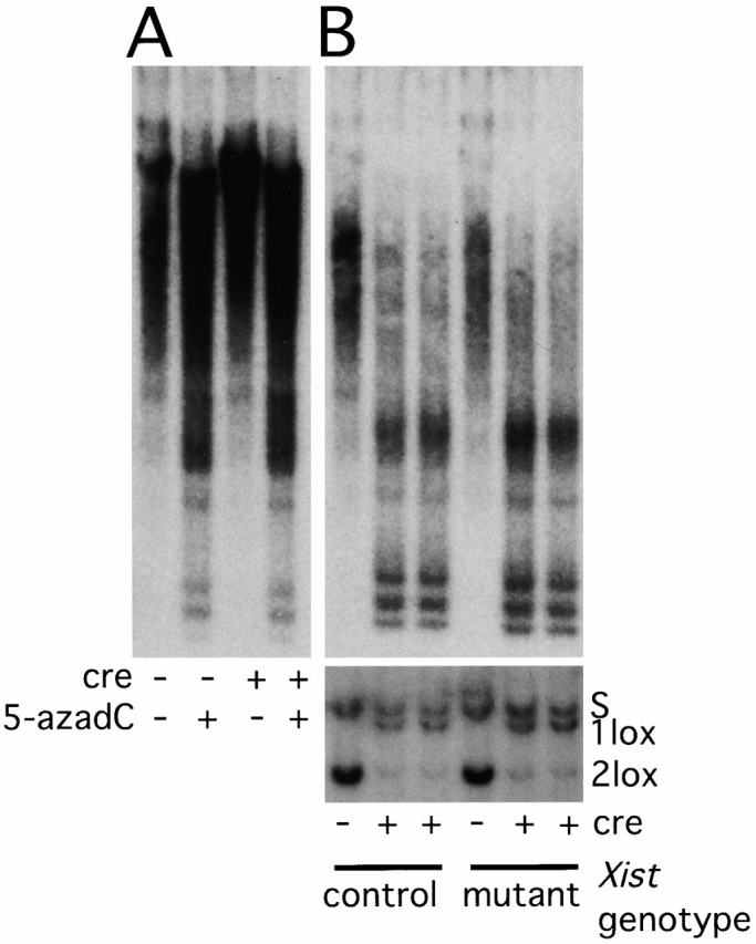
Xist RNA expression, methylation of CpG islands, and hypoacetylation of histone H4 are distinguishing features of inactive X chromatin. Here, we show that these silencing mechanisms act synergistically to maintain the inactive state. Xist RNA has been shown to be essential for initiation of X inactivation, but not required for maintenance. We have developed a system in which the reactivation frequency of individual X-linked genes can be assessed quantitatively. Using a conditional mutant Xist allele, we provide direct evidence for that loss of Xist RNA destabilizes the inactive state in somatic cells, leading to an increased reactivation frequency of an X-linked GFP transgene and of the endogenous hypoxanthine phosphoribosyl transferase (Hprt) gene in mouse embryonic fibroblasts. Demethylation of DNA, using 5-azadC or by introducing a mutation in Dnmt1, and inhibition of histone hypoacetylation using trichostatin A further increases reactivation in Xist mutant fibroblasts, indicating a synergistic interaction of X chromosome silencing mechanisms.


Figures

Figure 2
Figure 2
Reactivation of GFP in Xist mutant fibroblasts. FACS® analysis of cells. (A) Live cells were gated, and their GFP fluorescence was plotted against autofluorescence. Dots to the right of the diagonal represent cells in which GFP fluorescence is greater than autofluorescence and therefore are considered GFP positive. GFP-negative (+/Y) and GFP-positive (GFP/Y) populations are shown for control. Xist conditional mutant cell populations, before Cre-mediated deletion of Xist (−cre), contained a small number of GFP-positive cells. After Cre-mediated deletion of Xist (+cre), the number of GFP-positive cells increased. (B) Number of GFP-positive cells in primary conditional mutants and in controls with or without adenovirus-Cre infection on day 7 after infection. In Xist conditional mutants, Cre-mediated deletion of Xist led to a twofold increase in GFP-positive cells, whereas controls remained unchanged. (C) Long-term culture of SV-40 T-antigen–transformed conditional mutant cells. FACS® analysis was performed at various time points after adenovirus-Cre infection. Although initially we observed an increase in the number of GFP-positive cells after Cre-mediated deletion of Xist, the number decreased after another week in culture to reach the level of spontaneous reactivation and remained at that level for the duration of the experiment.
Figure 1
Figure 1
Generation of Xist conditional mutant fibroblasts with X-inactivated GFP and Hprt genes. (A) Map of the X chromosome with approximate genetic distances between genes. (B) Genotypes of Xist conditional mutant and control fibroblasts. Both cell types are phenotypically GFP negative and HAT sensitive, as the GFP transgene and the only functional Hprt allele are on the Xi and are inactivated (i). Conditional mutant cells carry the Xist2lox allele on Xi, whereas controls carry a wild-type Xist allele. (C) Cre-mediated deletion of Xist after adenovirus-Cre infection. Southern blotting of XbaI-digested DNA hybridized with probe 7 (pr7) indicates 100% recombination in primary (1°) and SV-40 T-antigen–immortalized (Tag) cells.
Figure 3
Figure 3
Reactivation of Hprt in Xist mutant fibroblasts. (A) HAT selection of Xist conditional mutant and control SV-40 T-antigen–immortalized fibroblasts with (+cre) or without (−cre) adenovirus-Cre infection. 2 × 10⁶ cells were plated, selected in HAT media, and then fixed and stained. Cells that reactivated the Xi-linked Hprt gene were able to proliferate and form colonies. No or very few colonies were observed on control plates and plates containing conditional mutants before adenovirus-Cre infection. However, after Cre-mediated deletion of Xist in conditional mutants, HAT-resistant colonies were routinely detected. (B) The proportion of HAT-resistant cells in the culture does not change significantly after culturing cells for 7 and 14 d, and 1 and 3 mo after Cre-mediated deletion of Xist.
Figure 4
Figure 4
FACS® analysis of Hprt-positive clones. The number of GFP-positive cells is higher in HAT-resistant clones than in bulk unselected cultures. Slow-growing clones reactivated GFP in a higher proportion of cells than fast-growing ones.
Figure 5
Figure 5
Late-replicating Xi chromosomes in clones with reactivated GFP and Hprt. (A) Mapping of the GFP transgene insertion site by DNA FISH. A Cy3-labeled GFP/Pgk-Puro probe (red) was hybridized to denatured chromosomes (DAPI, blue). Enlargement of a single X chromosome is shown with the centromere staining brighter with DAPI than the rest of the chromosome. The arrow shows the site of transgene integration near the centromere. The doublet signal corresponds to sister chromatids. (B) Analysis of replication timing of the inactive chromosome. BrdU incorporation into late-replicating regions of the genome was detected using a monoclonal anti-BrdU antibody and fluorescein-anti–mouse antibody (green) on DAPI-stained metaphase chromosome spreads (blue). Xi (arrow) was identified using the GFP/Pgk-Puro probe (red). In all clones analyzed, the Xi with reactivated genes was late replicating. (C) An enlargement of a single late-replicating Xi.
Figure 6
Figure 6
Synergism of X chromosome silencing mechanism. (A) FACS® analysis of GFP reactivation in Xist mutant fibroblasts with or without Cre infection treated with 5-azadC and/or TSA. TSA by itself had no effect. However, 5-azadC by itself, or followed by TSA treatment, led to significant GFP reactivation. Xist deleted cells were more sensitive to the treatment than those that did not delete Xist. (B) GFP reactivation in Xist/Dnmt1 double conditional mutant fibroblasts. Control cells (Dnmt12lox/Sand Xist +/Δ) delete Dnmt1 upon adenovirus-Cre infection, whereas Xist mutant cells (Dnmt12lox/Sand Xist2lox/ Δ) delete both Dnmt1 and Xist. Both primary (1°) and SV-40 T-antigen–immortalized (Tag) cells were analyzed. Deletion of Dnmt1 alone led to reactivation of GFP in ≤7% of primary and ≤15% of T-antigen–transformed cells. Deletion of Xist also in addition to Dnmt1 increased the number of GFP-positive cells about twofold. (C) The number of HAT-resistant colonies in Xist conditional mutant treated with 5-azadC. Cre-mediated deletion of Xist had a more significant effect on the number of HAT-resistant cells than 5-azadC treatment. However, deletion of Xist followed by 5-azadC treatment reactivated Hprt in a much higher proportion of cells than either treatment alone. Note the use of log scale.
Figure 7
Figure 7
Demethylation of genomic DNA. (A) Demethylation of genomic DNA after 5-azadC treatment of cells. Adenovirus-Cre–infected or –uninfected Xist conditional mutant cells were treated with 5-azadC or were left untreated. Genomic DNA was isolated and digested with the methylation-sensitive restriction enzyme HpaII. The blotted DNA was hybridized with an IAP probe to analyze demethylation of bulk genomic DNA. Demethylation due to 5-azadC treatment is indicated by the appearance of low molecular weight bands. (B) Adenovirus-Cre–mediated recombination in Dnmt12lox/S; Xist +/Δ (control) and Dnmt12lox/S; Xist2lox/ Δ (mutant) cells. Only Dnmt1 recombination in SV-40 T-antigen–immortalized cells is shown on Southern blots of SpeI-digested DNA hybridized with the HV probe (lower blot). Nearly 100% recombination is seen in both controls and mutants. The same genomic DNA samples were also digested with HpaII and hybridized with the IAP probe (upper blot). The appearance of more low molecular weight bands and the disappearance of high molecular weight bands indicate that the genomic DNA in these samples is more extensively demethylated than in 5-azadC–treated cultures.

Similar articles

Cited by

References

    1. Anton M., Graham F.L. Site-specific recombination mediated by an adenovirus vector expressing the Cre recombinase proteina molecular switch for control of gene expression. J. Virol. 1995;69:4600–4606. - PMC - PubMed
    1. Barr M.L., Carr D.H. Correlation between sex chromatin and chromosomes. Acta Cytol. 1962;6:34–45. - PubMed
    1. Borsani G., Tonlorenzi R., Simmler M.C., Dandolo L., Arnaud D., Capra V., Grompe M., Pizzuti A., Muzny D., Lawrence C. Characterization of a murine gene expressed from the inactive X chromosome. Nature. 1991;351:325–329. - PubMed
    1. Brockdorff N., Ashworth A., Kay G.F., Cooper P., Smith S., McCabe V.M., Norris D.P., Penny G.D., Patel D., Rastan S. Conservation of position and exclusive expression of mouse Xist from the inactive X chromosome. Nature. 1991;351:329–331. - PubMed
    1. Brockdorff N., Ashworth A., Kay G.F., McCabe V.M., Norris D.P., Cooper P.J., Swift S., Rastan S. The product of the mouse Xist gene is a 15-kb inactive X-specific transcript containing no conserved ORF and located in the nucleus. Cell. 1992;71:515–526. - PubMed

Publication types

MeSH terms