. 2001 May 15;29(10):2097-105.
doi: 10.1093/nar/29.10.2097.

Incoming nucleotide binds to Klenow ternary complex leading to stable physical sequestration of preceding dNTP on DNA

Affiliations

Incoming nucleotide binds to Klenow ternary complex leading to stable physical sequestration of preceding dNTP on DNA

S Ramanathan et al. Nucleic Acids Res. .

Abstract

Klenow-DNA complex is known to undergo a rate-limiting, protein conformational transition from an 'open' to 'closed' state, upon binding of the 'correct' dNTP at the active site. In the 'closed' state, Mg(2+) mediates a rapid chemical step involving nucleophilic displacement of pyrophosphate by the 3' hydroxyl of the primer terminus. The enzyme returns to the 'open' state upon the release of PPi and translocation permits the next round of reaction. To determine whether Klenow can translocate to the next site on the addition of the next dNTP, without the preceding chemical step, we studied the ternary complex (Klenow-DNA-dNTP) in the absence of Mg(2+). While the ternary complex is proficient in chemical addition of dNTPs in Mg(2+), as revealed by primer extensions, the same in Mg(2+)-deficient conditions lead to non-covalent (physical) sequestration of first two 'correct' dNTPs in the ternary complex. Moreover, the second dNTP traps the first one in the DNA-helix of the ternary complex. Such a dNTP-DNA complex is found to be stable even after the dissociation of KLENOW: This reveals the novel state of the dNTP-DNA complex where the complementary base is stacked in a DNA-helix non-covalently, without the phosphodiester linkage. Further, shuttling of the DNA between the polymerase and the exonuclease site mediates the release of such a DNA complex. Interestingly, Klenow in such a Mg(2+)-deficient ternary complex exhibits a 'closed' conformation.


Figures

Figure 1
Figure 1
Effect of next ‘correct’ nucleotide on the Klenow–DNA–dNTP ternary complex and the DNA complex: specificity and stability. (A) The ternary complex was formed with dATP (5 µM containing 10 µCi of [α-32P]dATP) as described in the Materials and Methods (lane 3). A parallel reaction also included unlabeled dTTP (50 µM) (second ‘correct’ nucleotide) (lane 4). The reaction corresponding to lane 4 was treated with 1% SDS prior to loading on the gel (lane 5). Other controls included: reaction described in lane 4 in the absence of either DNA substrate (lane 1) or Klenow (lane 2); 5′-32P-labeled oligomer duplex substrate marker (lane 6). Left, illustrative cartoon diagrams of ternary and DNA complexes are depicted, where the ellipsoid and the filled square represent Klenow and labeled dATP, respectively. (B) Ternary complex formed with Klenow, duplex DNA and dATP (5 µM containing 10 µCi of [α-32P]dATP) (lane 1) were pre-mixed with either dATP (lane 2), dTTP (lane 3), dGTP (lane 4) or dCTP (lane 5) (each at 50 µM) as unlabeled nucleotide. (C) Ternary and DNA complexes are formed by incubating duplex DNA, Klenow, [α-32P]dATP (5 µM) and dTTP (50 µM) either in the absence (lane 1) or presence (lane 4) of 10 mM Mg2+ as described in the Materials and Methods. The same reactions were treated with either 1% SDS (lanes 2 and 5) or with proteinase K (1 µg/µl) followed by an incubation at 25°C for 20 min (lanes 3 and 6) prior to loading on the gel.
Figure 1
Figure 1
Effect of next ‘correct’ nucleotide on the Klenow–DNA–dNTP ternary complex and the DNA complex: specificity and stability. (A) The ternary complex was formed with dATP (5 µM containing 10 µCi of [α-32P]dATP) as described in the Materials and Methods (lane 3). A parallel reaction also included unlabeled dTTP (50 µM) (second ‘correct’ nucleotide) (lane 4). The reaction corresponding to lane 4 was treated with 1% SDS prior to loading on the gel (lane 5). Other controls included: reaction described in lane 4 in the absence of either DNA substrate (lane 1) or Klenow (lane 2); 5′-32P-labeled oligomer duplex substrate marker (lane 6). Left, illustrative cartoon diagrams of ternary and DNA complexes are depicted, where the ellipsoid and the filled square represent Klenow and labeled dATP, respectively. (B) Ternary complex formed with Klenow, duplex DNA and dATP (5 µM containing 10 µCi of [α-32P]dATP) (lane 1) were pre-mixed with either dATP (lane 2), dTTP (lane 3), dGTP (lane 4) or dCTP (lane 5) (each at 50 µM) as unlabeled nucleotide. (C) Ternary and DNA complexes are formed by incubating duplex DNA, Klenow, [α-32P]dATP (5 µM) and dTTP (50 µM) either in the absence (lane 1) or presence (lane 4) of 10 mM Mg2+ as described in the Materials and Methods. The same reactions were treated with either 1% SDS (lanes 2 and 5) or with proteinase K (1 µg/µl) followed by an incubation at 25°C for 20 min (lanes 3 and 6) prior to loading on the gel.
Figure 2
Figure 2
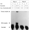
Second ‘correct’ nucleotide is part of the ternary complex, but not the DNA complex. Ternary complexes were formed by incubating Oligo A with Klenow and [α-32P]dATP (5 µM) (lane 1). In a parallel reaction, additionally, dTTP (10 µM) was added (lane 2). Similarly, ternary complexes were formed by incubating Oligo B with Klenow and [α-32P]dATP (10 µM) (lane 3) to which, additionally, dTTP (5 µM) was added (lane 4). All the samples were analyzed by native PAGE as described in the text.
Figure 3
Figure 3
The relationship between DNA complexes and the exonuclease domain in the polymerase. Ternary complexes were formed with a polymerase that is either exonuclease proficient (Klenow polymerase) or deficient (Klenow exo–) in the presence of [α-32P]dATP (5 µM) and dTTP (50 µM) without Mg2+ (see Materials and Methods), followed by native PAGE.
Figure 4
Figure 4
Effect of dTTP and dCTP concentration on ternary and DNA complex. (A) Ternary complexes were formed by incubating duplex DNA with Klenow, [α-32P]dATP (5 µM) and increasing concentrations of dTTP (10–400 µM), followed by native PAGE. (B) Ternary complexes formed by incubating duplex DNA with Klenow, [α-32P]dATP (5 µM) and dTTP (50 µM) were challenged with increasing concentration of dCTP (0–66 µM) at room temperature for 5 min, followed by native PAGE. Radioactivity associated with ternary complexes, DNA complexes and free label were quantitated using a phosphorimager. Radioactivity associated with ternary (circles) and DNA complexes (triangles) were expressed as percentage of total radioactivity and the mean of three independent experiments is plotted as a function of dTTP (A) and dCTP (B) concentration.
Figure 4
Figure 4
Effect of dTTP and dCTP concentration on ternary and DNA complex. (A) Ternary complexes were formed by incubating duplex DNA with Klenow, [α-32P]dATP (5 µM) and increasing concentrations of dTTP (10–400 µM), followed by native PAGE. (B) Ternary complexes formed by incubating duplex DNA with Klenow, [α-32P]dATP (5 µM) and dTTP (50 µM) were challenged with increasing concentration of dCTP (0–66 µM) at room temperature for 5 min, followed by native PAGE. Radioactivity associated with ternary complexes, DNA complexes and free label were quantitated using a phosphorimager. Radioactivity associated with ternary (circles) and DNA complexes (triangles) were expressed as percentage of total radioactivity and the mean of three independent experiments is plotted as a function of dTTP (A) and dCTP (B) concentration.
Figure 5
Figure 5
Kd (DNA) analyses of Klenow–DNA complexes (binary and ternary) formed in the absence of Mg2+. Binary complexes were formed by incubating 5′ end-labeled duplex substrate (5 nM) with increasing amounts of Klenow (0–2400 nM) in the reaction buffer [50 mM Tris–HCl (pH 7.5), 0.1 mM EDTA, 1 mM DTT] on ice for 15 min, followed by analyses on native 8% polyacrylamide gel at 4°C. The gel was dried and autoradiographed. Similar gel-shift analyses were carried out for ternary complexes formed by incubating 5′ end-labeled duplex substrate (5 nM) with increasing amounts of Klenow (0–2400 nM) in the presence of dATP (0.5 mM). Radioactivity associated with gel-shifted binary/ternary complexes and free DNA was quantified using a phosphorimager. Percentage of total radioactivity associated with the binary/ternary complexes (indicated as percentage DNA complexed) was plotted against Klenow concentration. The data points represent a mean of three independent experiments. The binding isotherms were fitted using Sigma-plot program based on which Kd was computed. Binary complex (circle): ternary complex (square).
Figure 6
Figure 6
Analyses of ‘open’ and ‘closed’ Klenow complexes by trypsin fingerprinting assay. Klenow complexes were formed in the presence of the specified nucleotide, followed by trypsin fingerprinting of Klenow (see the Materials and Methods). The samples were analyzed on 10% SDS–polyacrylamide gel, where the fragments were visualized by silver staining.

Similar articles

Cited by

References

    1. Bryant F.R., Johnson,K.A. and Benkovic,S.J. (1983) Elementary steps in the DNA polymerase I reaction pathway. Biochemistry, 22, 3537–3546. - PubMed
    1. Mizrahi V., Henrie,R.N., Marlier,J.F., Johnson,K.A. and Benkovic,S.J. (1985) Rate limiting steps in the DNA polymerase I reaction pathway. Biochemistry, 24, 4010–4018. - PubMed
    1. Kuchta R.D., Mizrahi,V., Benkovic,P.A., Johnson,K.A. and Benkovic,S.J. (1987) Kinetic mechanism of DNA polymerase I (Klenow). Biochemistry, 26, 8410–8417. - PubMed
    1. Kuchta R.D., Benkovic,P.A. and Benkovic,S.J. (1988) Kinetic mechanism whereby DNA polymerase I (Klenow) replicates DNA with high fidelity. Biochemistry, 27, 6716–6725. - PubMed
    1. Eger B.T., Kuchta,R.D., Carroll,S.S., Benkovic,P.A., Dahlberg,M.E., Joyce,C.M. and Benkovic,S.J. (1991) Mechanism of DNA replication fidelity for the three mutants of DNA polymerase I: Klenow fragment KF (exo+), KF (polA5), and KF (exo–). Biochemistry, 30, 1441–1448. - PubMed

MeSH terms