Comparative Study
. 2001 May 15;29(10):2117-26.
doi: 10.1093/nar/29.10.2117.

A reliable assessment of 8-oxo-2-deoxyguanosine levels in nuclear and mitochondrial DNA using the sodium iodide method to isolate DNA

Affiliations
Comparative Study

A reliable assessment of 8-oxo-2-deoxyguanosine levels in nuclear and mitochondrial DNA using the sodium iodide method to isolate DNA

M L Hamilton et al. Nucleic Acids Res. .

Abstract

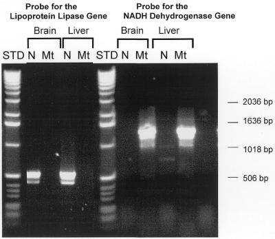
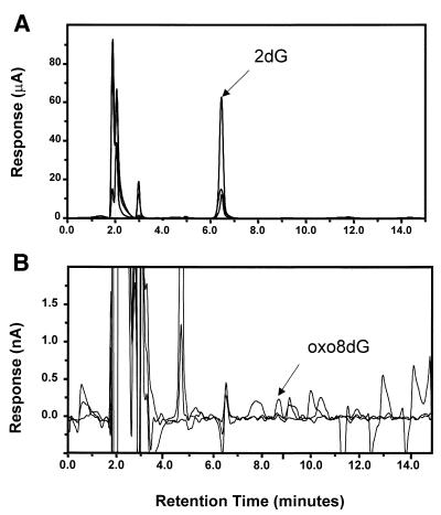
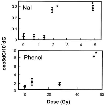
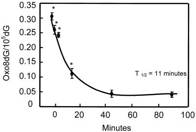
A major controversy in the area of DNA biochemistry concerns the actual in vivo levels of oxidative damage in DNA. We show here that 8-oxo-2-deoxyguanosine (oxo8dG) generation during DNA isolation is eliminated using the sodium iodide (NaI) isolation method and that the level of oxo8dG in nuclear DNA (nDNA) is almost one-hundredth of the level obtained using the classical phenol method. We found using NaI that the ratio of oxo8dG/10(5 )deoxyguanosine (dG) in nDNA isolated from mouse tissues ranged from 0.032 +/- 0.002 for liver to 0.015 +/- 0.003 for brain. We observed a significant increase (10-fold) in oxo8dG in nDNA isolated from liver tissue after 2 Gy of gamma-irradiation when NaI was used to isolate DNA. The turnover of oxo8dG in nDNA was rapid, e.g. disappearance of oxo8dG in the mouse liver in vivo after gamma-irradiation had a half-life of 11 min. The levels of oxo8dG in mitochondrial DNA isolated from liver, heart and brain were 6-, 16- and 23-fold higher than nDNA from these tissues. Thus, our results showed that the steady-state levels of oxo8dG in mouse tissues range from 180 to 360 lesions in the nuclear genome and from one to two lesions in 100 mitochondrial genomes.


Figures

Figure 1
Figure 1
Characterization of nDNA and mtDNA isolated from liver and brain. nDNA and mtDNA isolated from liver and brain by the NaI method were used as PCR templates for primers to the NADH dehydrogenase gene (found in the mtDNA) and the lipoprotein lipase gene (found in the nDNA). A 1400 bp product was generated with mtDNA using primers for the NADH dehydrogenase gene, and a 500 bp product was generated with nDNA using primers for the lipoprotein lipase gene. A 1 kb DNA Ladder (Gibco/BRL, Santa Fe, NM) is shown as a standard.
Figure 2
Figure 2
Analysis of DNA hydrolysates by HPLC-EC. nDNA (50 µg) isolated from liver by the NaI method was analyzed. (A) The retention time of 2dG was ∼6.5 min with potential settings across three channels of 550, 650 and 750 mV. (B) The retention time for oxo8dG was ∼8.7 min with potential settings across three channels of 250, 350 and 450 mV. The peaks for 2dG and oxo8dG were identified based on the co-injection of authentic standards as described in the Materials and Methods.
Figure 3
Figure 3
Effect of whole-body γ-irradiation on the ratio of oxo8dG/105 2dG in nDNA from mouse liver. Mice were exposed to acute whole-body γ-irradiation at doses of 0–50 Gy. Following irradiation, the mice were immediately killed and the liver divided in half. nDNA was isolated from the two halves of the liver using either the phenol or NaI method. Each value is expressed as a mean ± SEM of data from six mice. The data were analyzed statistically using a one-way ANOVA with a Dunnett’s test and the values shown by the asterisks are significantly higher than the control values (no γ-irradiation).
Figure 4
Figure 4
Time course for the removal of oxo8dG from the nDNA of liver tissue following acute whole-body γ-irradiation. Mice were exposed to 2 Gy of whole-body γ-irradiation and euthanized at 0, 2, 5, 20, 45 and 90 min after irradiation. The nDNA was isolated from the liver using the NaI method. The ratio of oxo8dG/105dG represents the mean ± SEM of data collected from six mice. The data were statistically analyzed using a one-way ANOVA and the values shown by the asterisks are significantly higher (P < 0.05) than the control value (no γ-irradiation).

References

    1. Beckman K.B. and Ames,B.N. (1999) Endogenous oxidative damage of mtDNA. Mutat. Res., 424, 51–58. - PubMed
    1. Ames B.N. and Gold,L.S. (1991) Endogenous mutagens and the causes of aging and cancer. Mutat. Res., 250, 3–16. - PubMed
    1. Wagner J.R., Hu,C.C. and Ames,B.N. (1992) Endogenous oxidative damage of deoxycytidine in DNA. Proc. Natl Acad. Sci. USA, 89, 3380–3384. - PMC - PubMed
    1. Dizdaroglu M. (1998) Facts about the artifacts in the measurement of oxidative DNA base damage by gas chromatography-mass spectrometry. Free Radic. Res., 29, 551–563. - PubMed
    1. Kaur H. and Halliwell,B. (1996) Measurement of oxidized and methylated DNA bases by HPLC with electrochemical detection. Biochem. J., 318, 21–23. - PMC - PubMed

Publication types

MeSH terms